|
|
|
|
|
|
|
|
|
|
|
|
|
||
|
|
|
||
D Implant for Lateral?
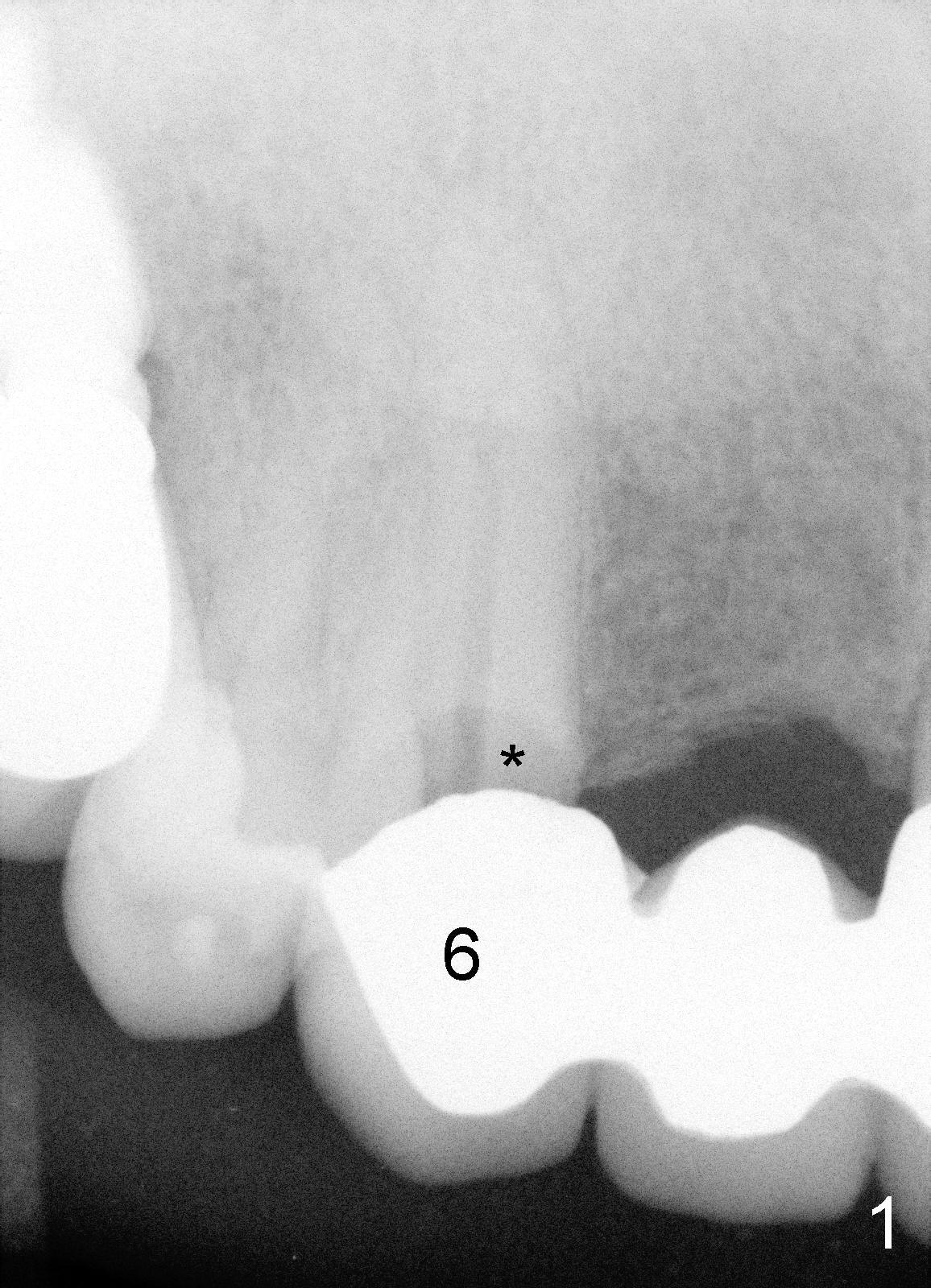
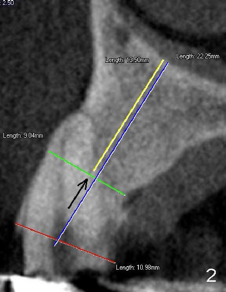
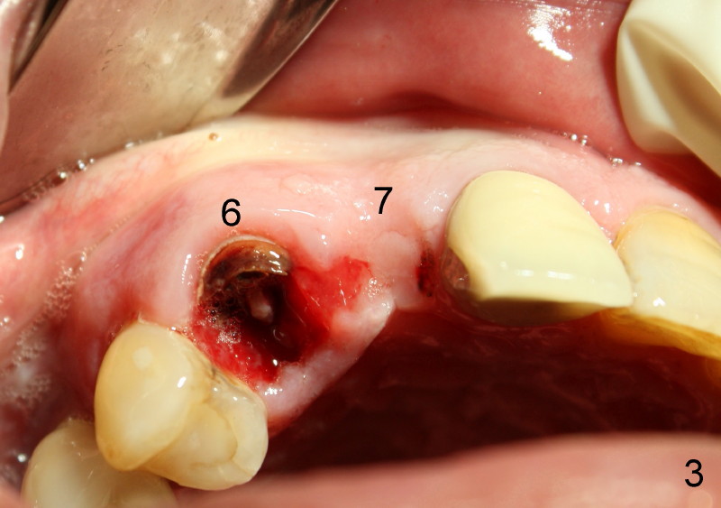
A 73-year-old man feels that upper right bridge is loose. There is subgingival caries (*) under one of its retainers (#6 in Fig.1). CBCT shows that bone volume is sufficient for immediate implant at #6 (Fig.2). When the bridge is sectioned between the pontic #7 and retainer #8, the pontic #7 and retainer #6 complex falls out by themselves. The tooth #6 is deemed nonsalvageable (albeit vital); the labial plate of #7 is atrophic (Fig.3).
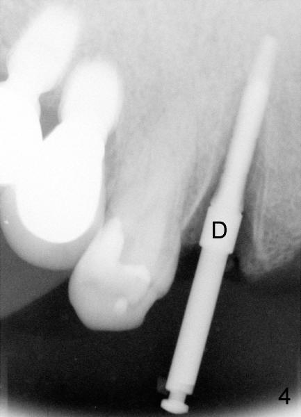
Immediately after atraumatic extraction of #6 residual root, the socket is irrigated with normal saline and Chlorhexidine. There is no granulation tissue in the apical area. The root stump is 6 mm mesiodistally and 8.5 mm labiopalatally. Tatum 2 mm pilot drill is used with copious irrigation to penetrate the palatal plate of the socket (as shown by arrow in Fig.2) at the depth of 20 mm from gingival margin. Bicon reamers (cylindrical, from 2.5 mm to 4.0 mm in a sequential order) are used (with 400:1 reduction) for osteotomy without irrigation. Autogenous bone graft is collected to fill the gap between implant and socket. Fig.4 shows 3.0 mm reamer in place (20 mm from the gingival margin). Finally Tatum tapered implant 6x20 mm is torqued in with primary stability (Fig.5: I). There is no gap between the implant and the socket except palatally, where the bone graft is inserted. Two release incisions are placed palatally. The flap is raised. The periosteum is scored at the base. The palatal flap is advanced labially to close the palatal gap with 4-0 chromic gut suture.
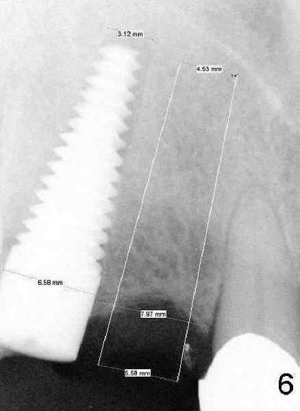
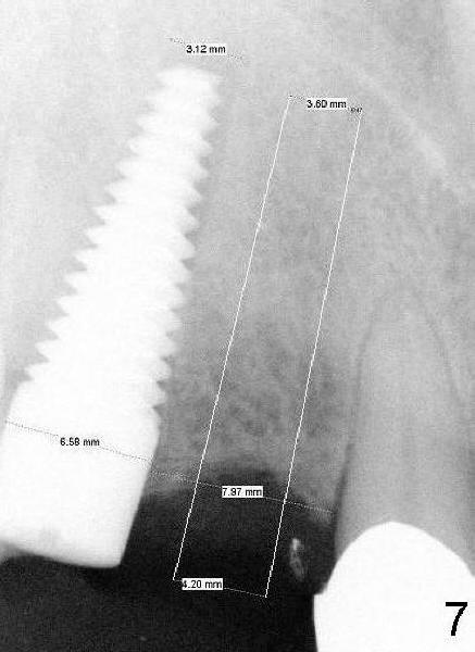
There is approximately 8 mm between the tooth #8 and the implant at the site of #6 (Fig.6,7). The distance appears to be enough to place D2 and D1 implants (Fig.6,7, respectively).
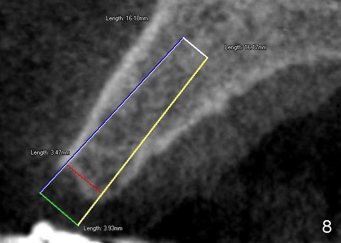
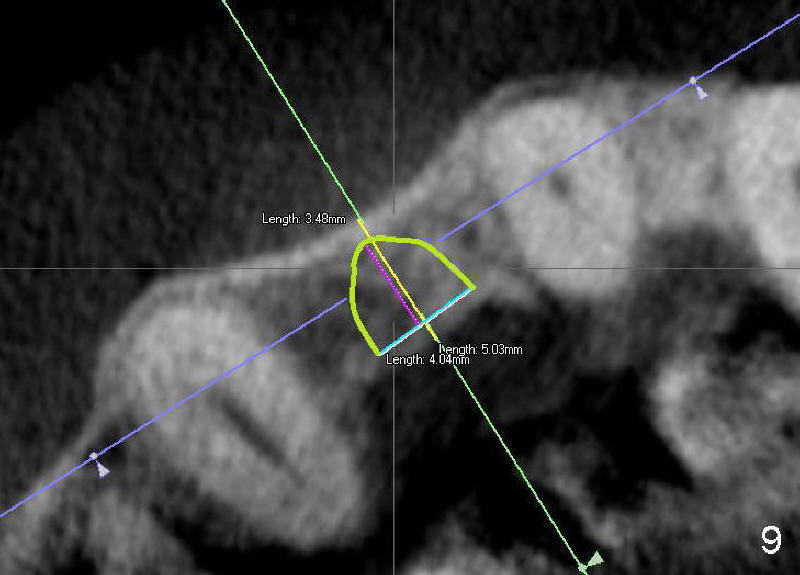
According to pre-op CBCT analysis, D1 short implant can be safely placed at the site of #7 (Fig.8,9, as compared to Fig.3). Which of D implants is the most appropriate?
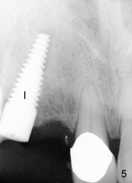
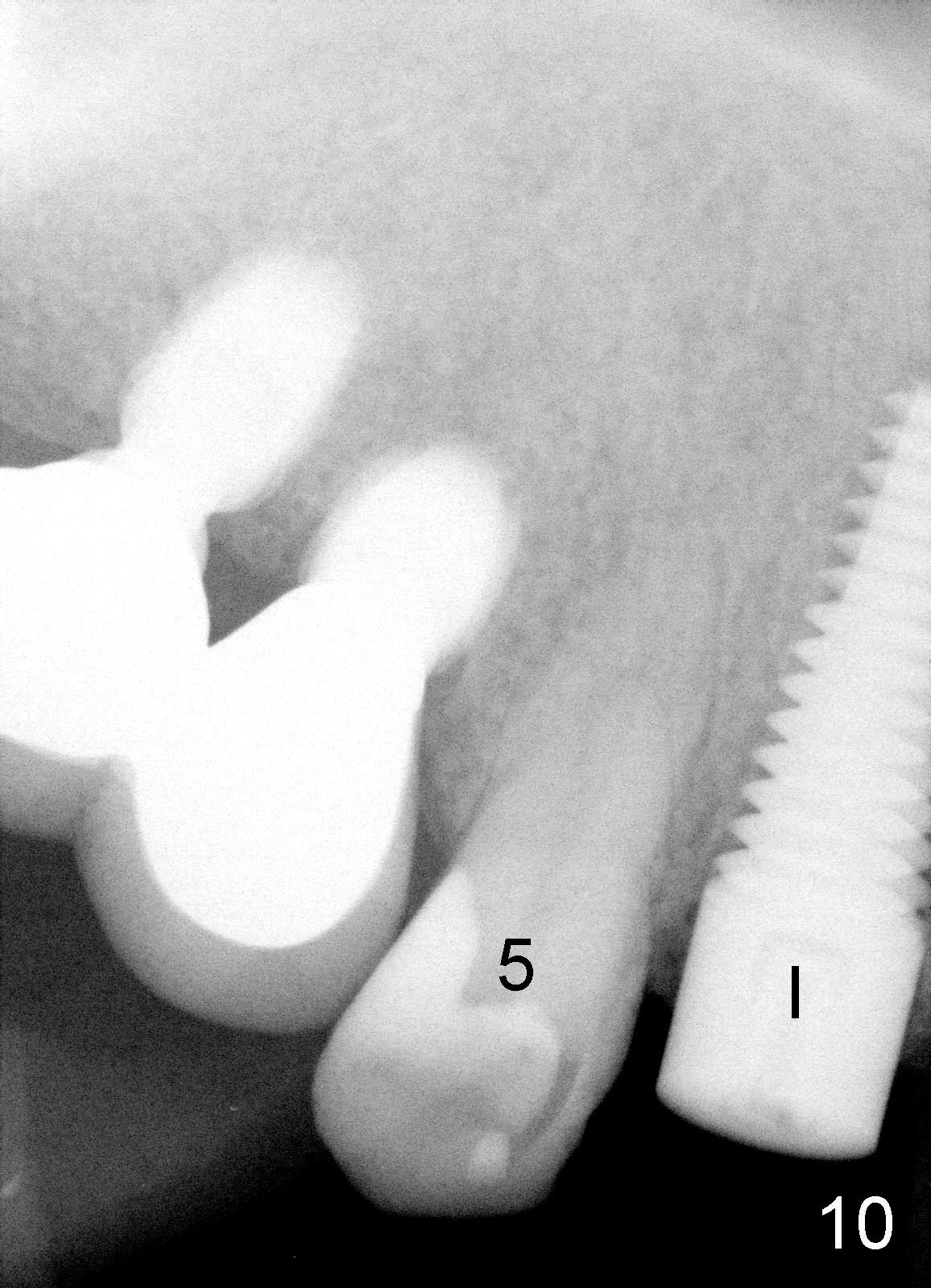
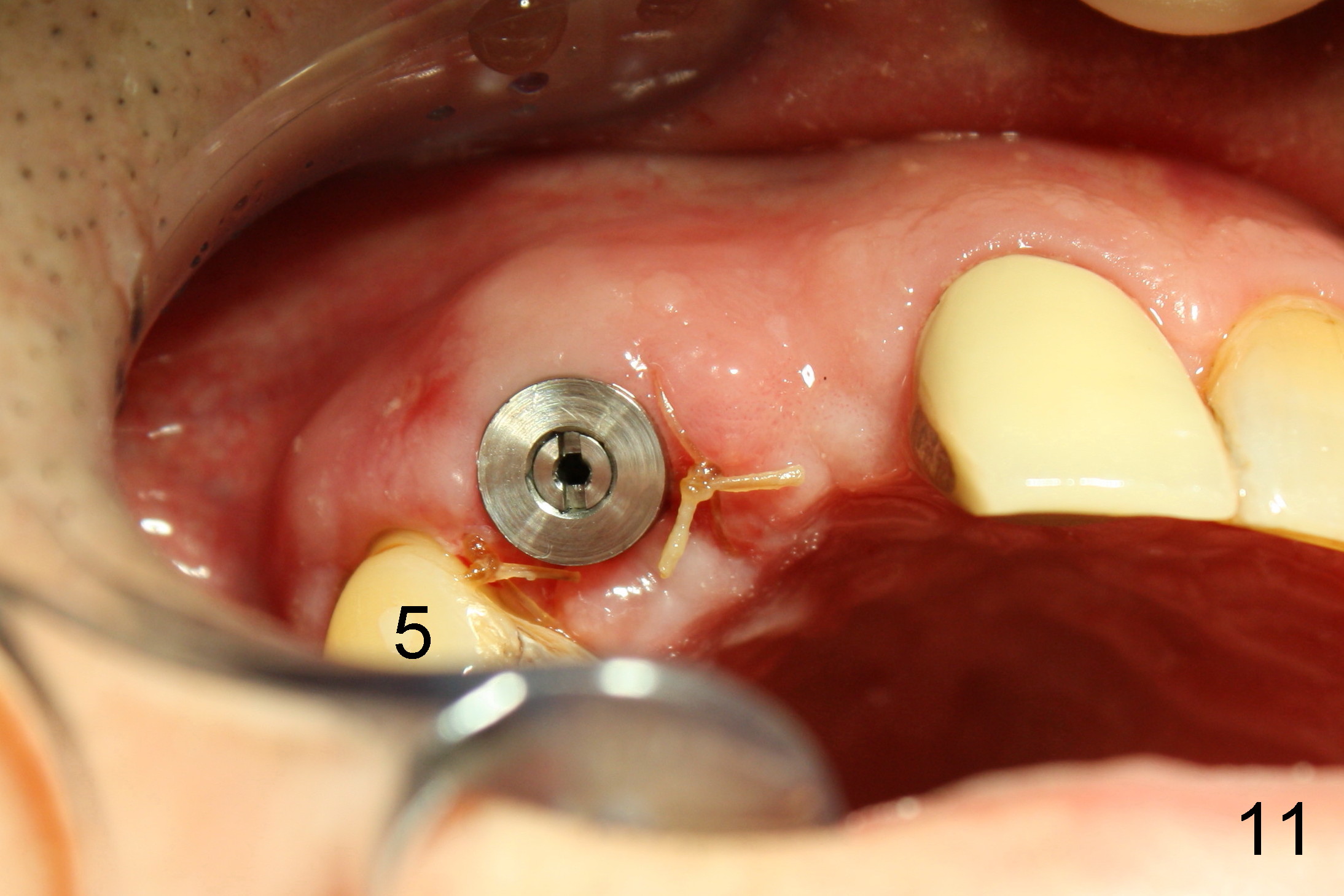
One week after #6 immediate implant placement, the patient is doing well. There is apparently enough clearance between the tooth #5 and the implant in term of bone (Fig.10, compare to Fig.4) and soft tissue (Fig.11, compare to Fig.3).
Xin Wei, DDS, PhD, MS 1st edition 12/08/2011, last revision 12/14/2011