


|
|
|
|
|
|
|
|
|
|
|
|
|
||
牙冠口扫
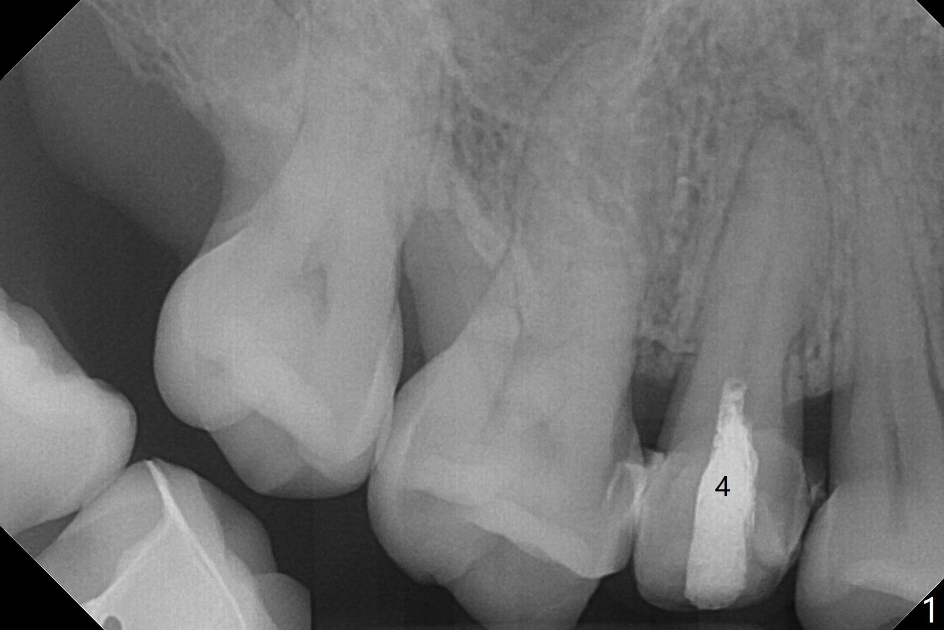
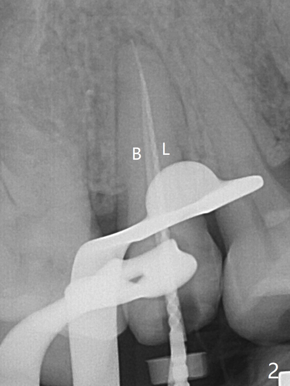
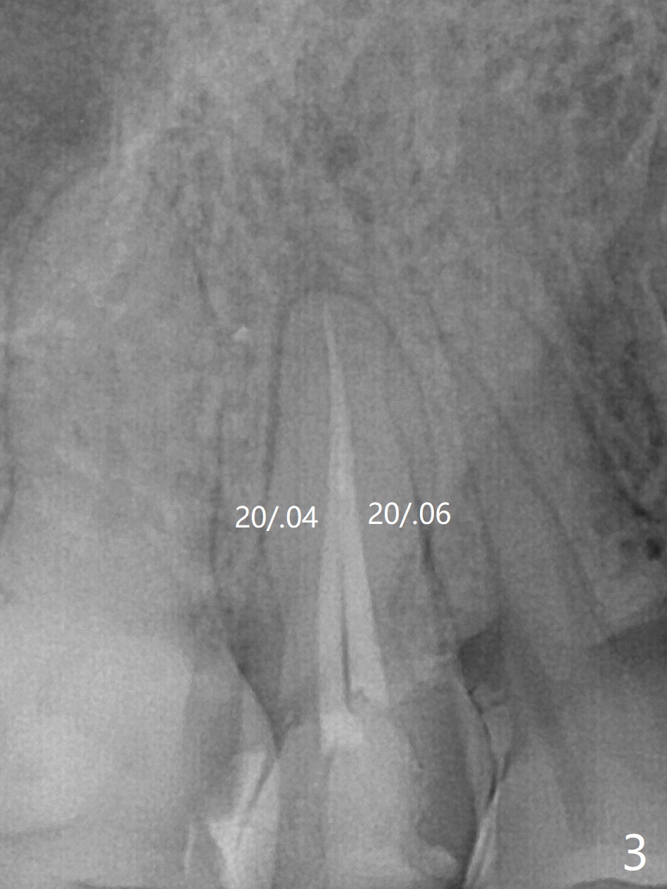
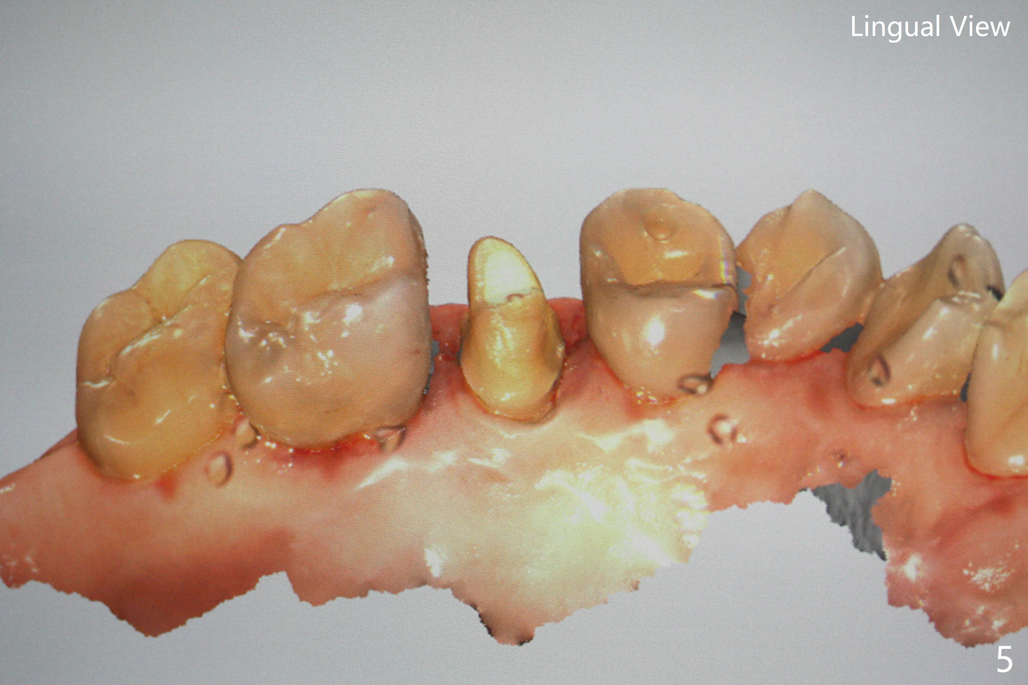
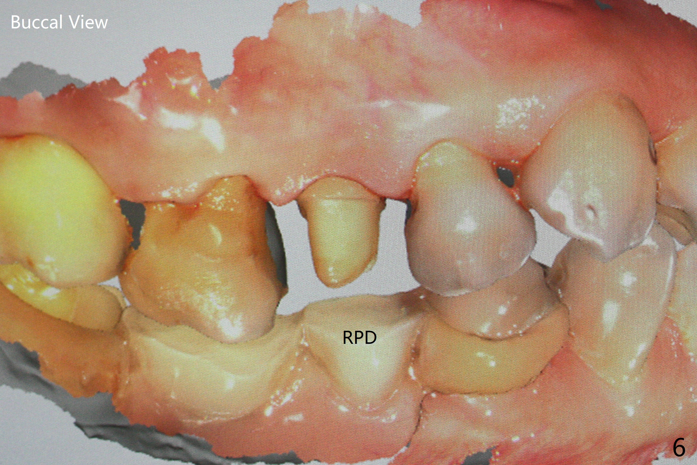
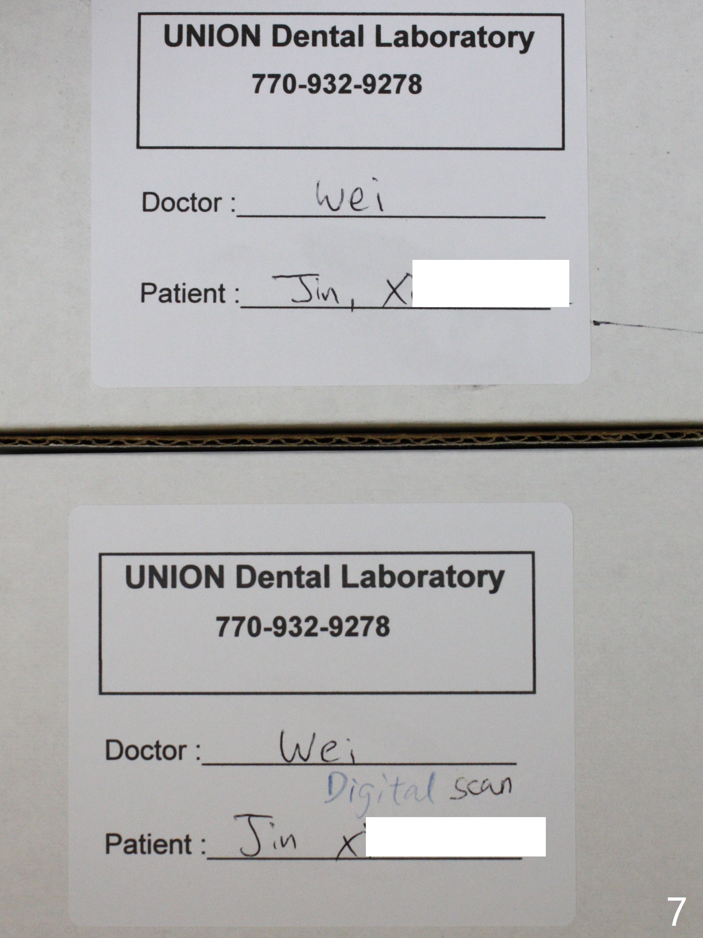
52岁女去年在外州开始4号牙根管治疗(图一),由于新冠病毒和搬家而拖延治疗,现在要求完成根管治疗(图二,三),B: 颊侧(主牙胶尖:20/.04);L: 舌侧(旋转锉:20/.04)。Shining口扫(图四(咬合面观),五(舌侧观),六(颊侧观;对合:局部托牙 (RPD)))。为了保险起见,要求实验室制作两个牙冠:取模,口扫。帮助实验室建立完善数字化系统,从而帮助临床工作。备牙边缘清晰(图八)。牙冠边缘与数字模型(图九)和牙齿吻合,天衣无缝。
Shining Oral Scanner and Crown Prep Margin
Root canal therapy (RCT) was initiated for the tooth #4 of a 52-year-old lady out of state approximately 8 months earlier (Fig.1). The patient requested finishing the treatment and permanent crown fabrication. The buccal (Fig.2 B (master cone 20/.04)) and lingual (L (rotary file 20/.04))) canals fuse near the apex. RCT was done with insertion of 20/.04 and 20/.06 master cones in the buccal and lingual canals, respectively, followed by composite build-up (Fig.3). With basically shoulder margin (not feather margin, chamfer margin ok), it is easy to scan (Fig.4-6 (RPD: removable partial denture)). Return to Oral Scanner Xin Wei, DDS, PhD, MS 1st edition 06/05/2021, last revision 06/25/2021