|
|
|
|
|
Food Impaction
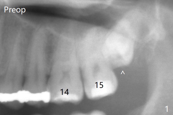
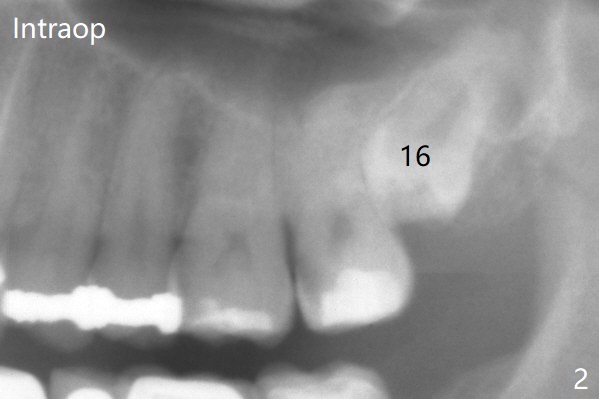
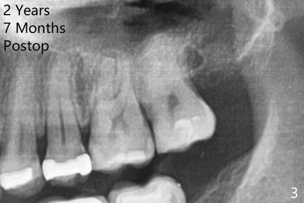
A 37-year-old man complains of food impaction between #14 and 15. Panoramic X-ray shows mesioangular impaction of #16 (Fig.1 ^), which may press #15 root to rotate the tooth so that there is a diastema between #14 and 15. When the impacted tooth is removed, it looks like a microdontia. Intraop X-ray reveals a normal sized 3rd molar in situ (Fig.2: 16). Therefore the microdontia removed is a supernumerary tooth. The tooth #16 is then extracted. The diastema appears to persist 2 years 7 months postop (Fig.3), although gingival swelling and pain reduces. The follow up panoramic X-ray reveals a significant increase in PARL at #20. Return to Professionals Xin Wei, DDS, PhD, MS 1st edition 05/14/2020, last revision 05/16/2020