|
|
|
|
|
|
|
|
|
|
|
|
Journey to Find 4th Canal in a Lower Molar
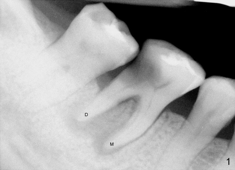
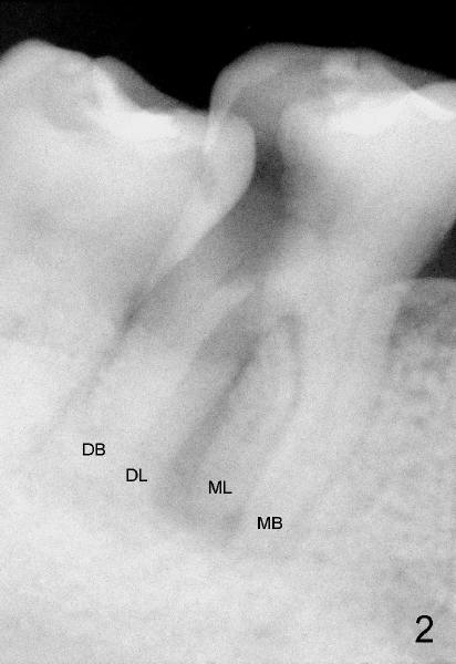
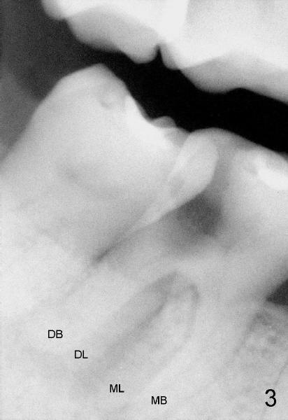
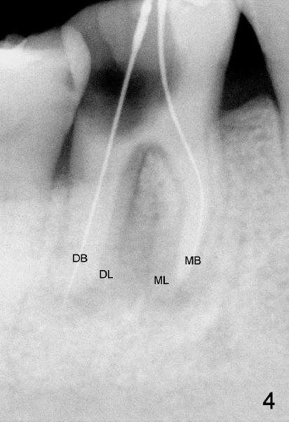
A healthy 43-year-old lady requests saving the tooth #30 with large DO caries and perioapical radiolucency (Fig.1). The tooth appears to have mesial (M) and distal (D) roots. Different angulation PA (Fig.2) and Bitewing (Fig.3) show that there are totally 4 roots/canals, which is not noted by the operator before RCT or after initiation of RCT (Fig.4). The distal canal orifice is not in the center. The distolingual (DL) canal orifice is searched without success. MB, ML, and D canals are debrided with rotary file 30/.04 at working lengths (WL).
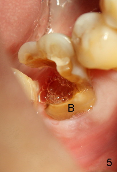
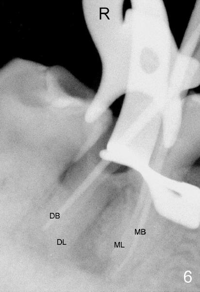
On the second appointment, the buccal wall is fractured (Fig.5: B). Visibility improves. Straight access is obtained, particularly mesially, but DL orifice is still not found. So D canal appears to be the only canal distally. It is further debrided with 40/.04 file. PA is taken when master cones are inserted (Fig.6). This time DL root is quite obvious.
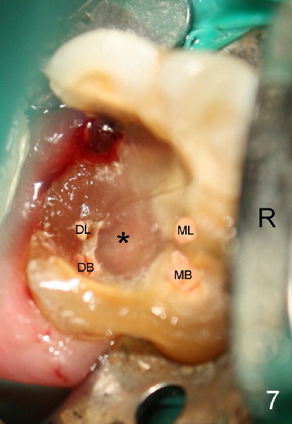
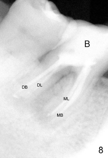
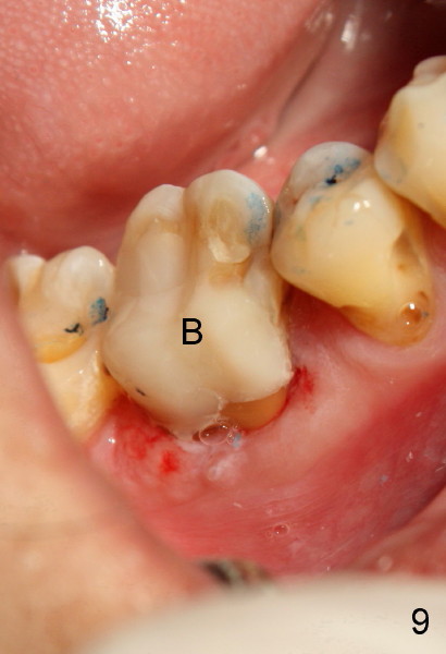
To further improve visibility, the arm of rubber dam clamp is changed from distal to mesial aspect of #30 (compare R in Fig.6 and 7). DL orifice is finally found, close to DB one. The DL canal is obliterated. It is debrided until #20 hand file short of WL. Fig.7 shows the chamber after RCT with gutta percha. * indicates thin pulpal floor. Fig.8 and 9 are taken after build-up (B). It appears that distal canals are divergent apically and that there are most likely two distal roots.
Xin Wei, DDS, PhD, MS 1st edition 03/1/2012, last revision 03/14/2012