|
|
|
|
|
|
|
Simultaneous Bone Expansion and Sinus Lift
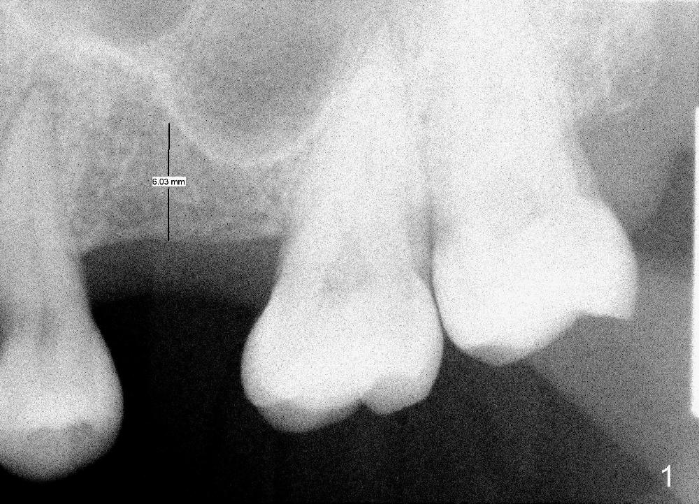
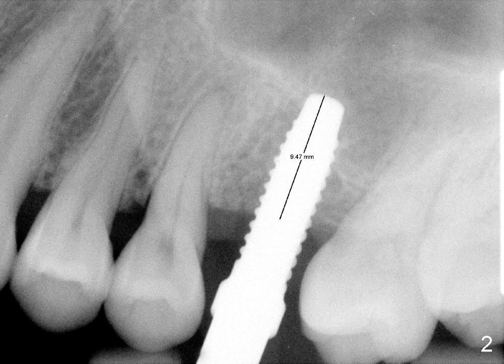
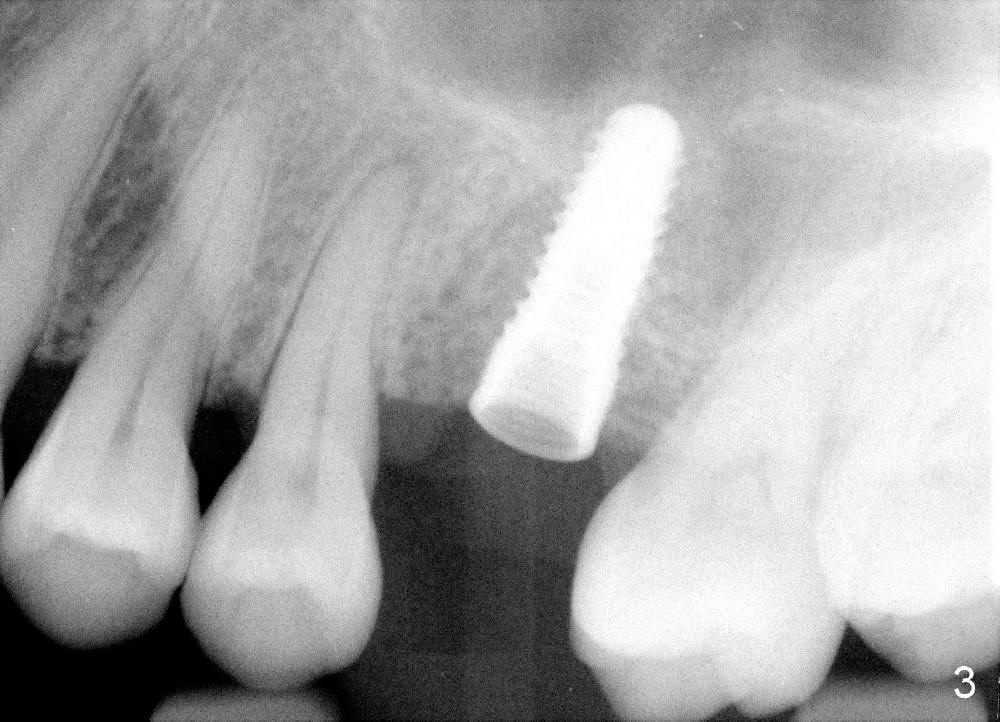
After local anestheisa, an incision is made. The initial osteotomy at the site of the upper left 1st molar is created by 1.6 mm pilot drill at the depth of 6 mm (Fig.1), followed by insertion of bone expanders 2.6, 3.0, and 3.4 mm at approximately 7, 8, and 9 mm deep (Fig.2). A 4.1 mm bone tap is inserted approximately 10 mm without much binding. Mineralized allograft mixed with Osteogen is used for sinus lift. A 4.5x12 mm implant is placed with insertion torque around 35 Ncm (Fig.3). A 5.2 mm healing abutment is placed. The flaps are sutured. The wound is covered by perio dressing.
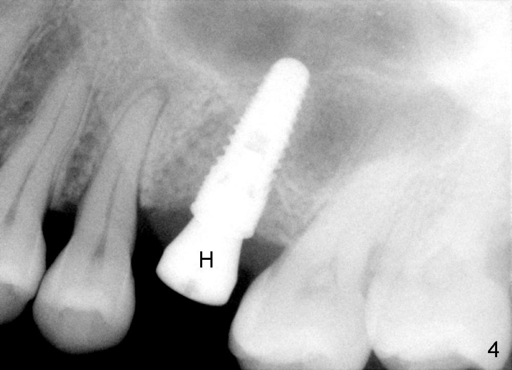
Fig.4 is taken 3.5 months postop. There is some bone surrounding the apex of the implant.
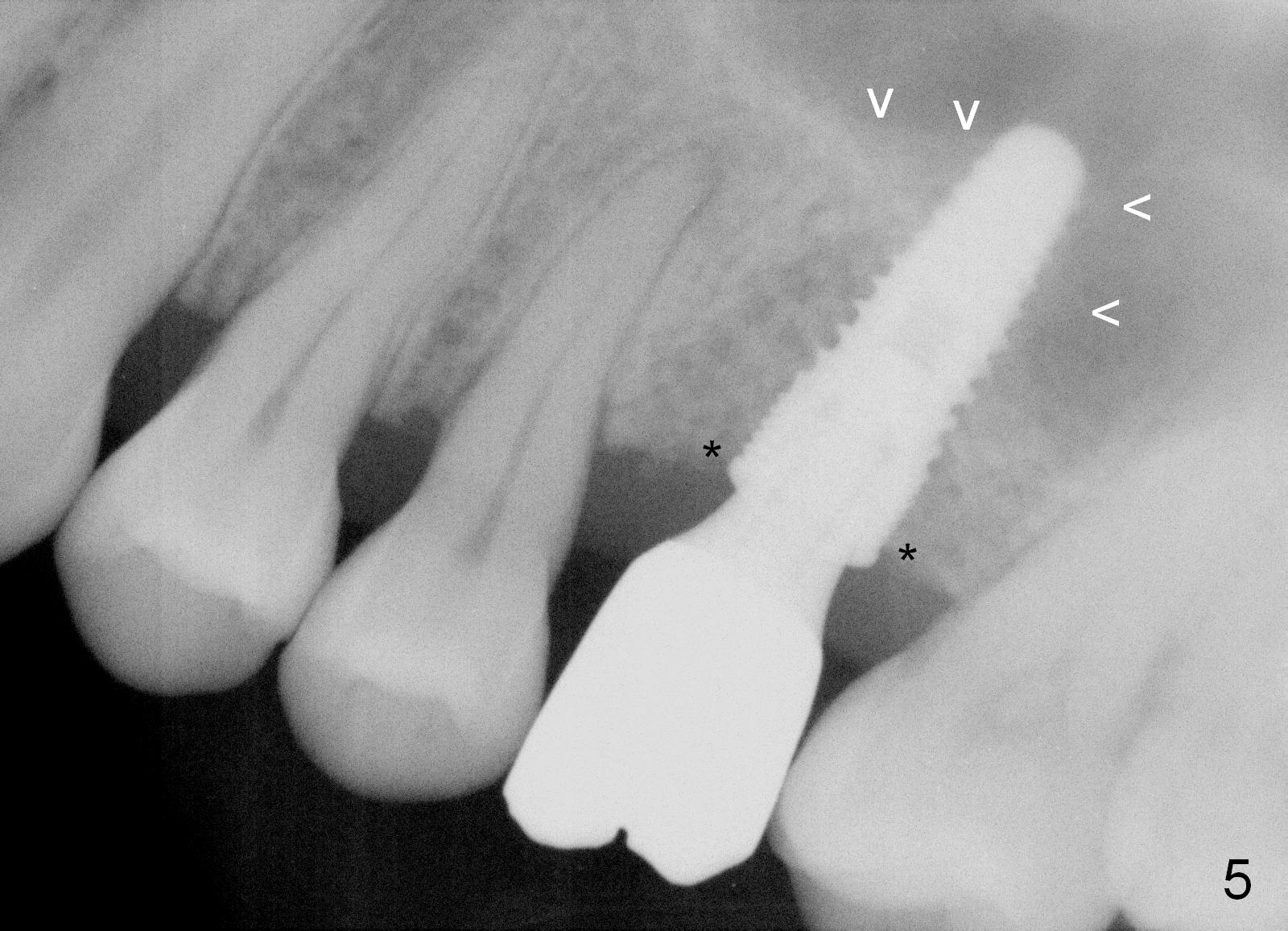
The patient returns for follow up 2.5 months post cementation. There is no crestal bone resorption (Fig.5 *), while the bone still surrounds the apex of the implant (arrowheads).
Return to Sinus Lift,
Professionals,
Dr. Wu
Xin Wei, DDS, PhD, MS 1st edition 07/10/2014, last revision 04/13/2015