
|
|
|
|
|
|
|
|
|
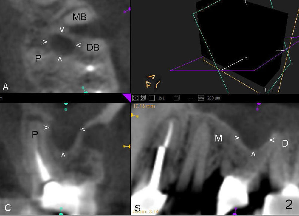
Coronal Extension of the Sinus Floor
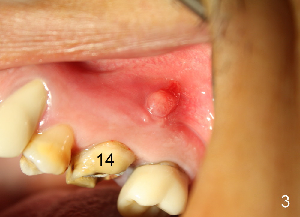
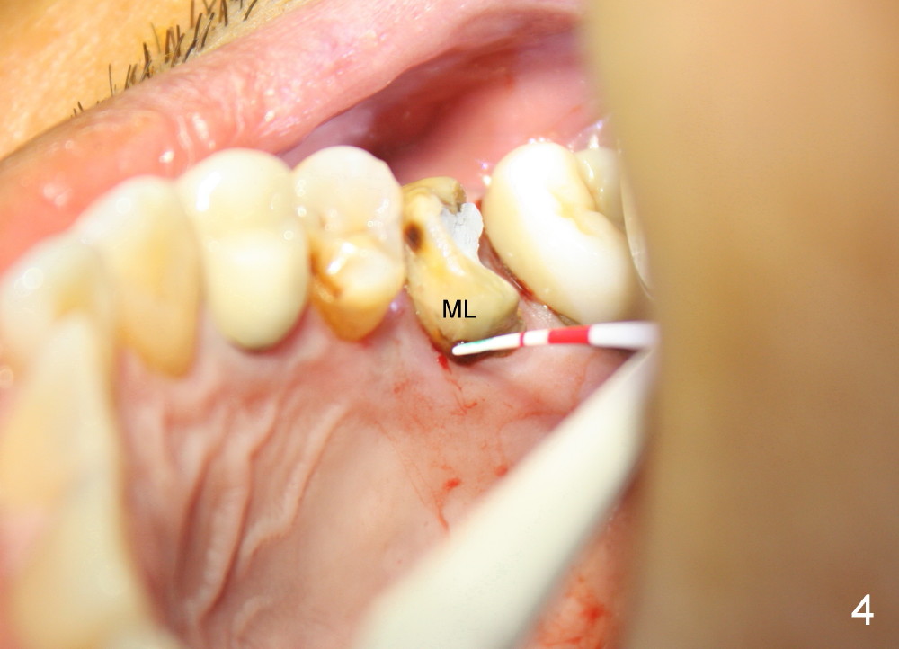
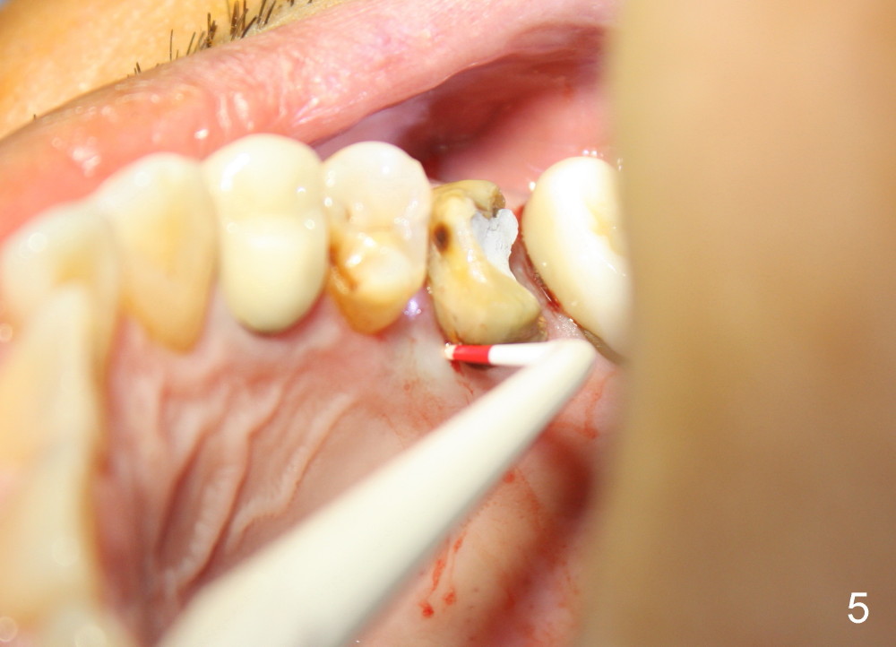
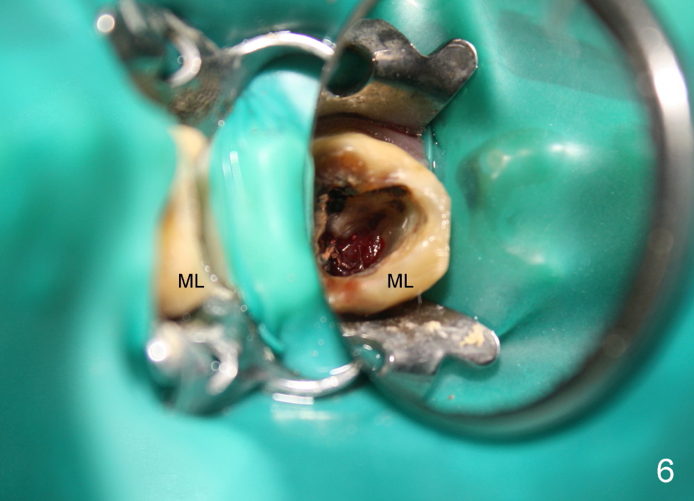
A 49-year-old man has multiple restoration (Fig.1). The tooth #14 appears to be non salvageable (Fig.2-6). The most interesting observation is that the sinus floor (Fig.1 ^) appears to be coronal to the tips of all three roots (mesiobuccal (MB), distobuccal (DB) and palatal (P)). This anatomic feature is confirmed by CBCT study (Fig.2). It will increase insertion torque of an implant to be placed if the sinus floor is raised among the root tips. That is, the apical portion of a large implant is engaged to the area among the apical portion of the three sockets. Let us use Fig.2 Coronal section for design of immediate implant with sinus lift. Click each figure for narrative.
Return to Sinus Lift
Xin Wei, DDS, PhD, MS 1st edition 05/18/2014, last revision 05/18/2014