 |
 |
 |
|
Fig.1 |
Fig.2 |
Fig.3 |
Dental Education Lecture: Which Tooth is Implant Supported? (2nd page)
|
|
|
|
|
Fig.1 |
Fig.2 |
Fig.3 |
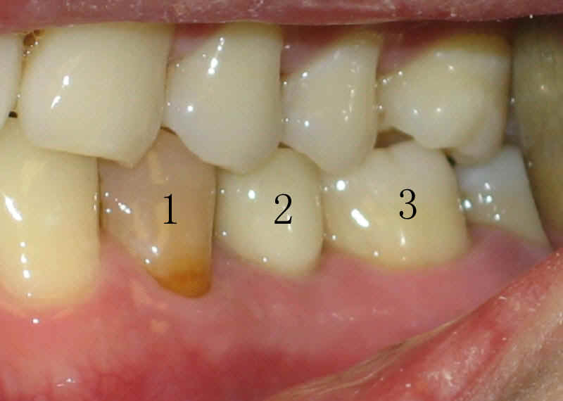
In the main page of this lecture, we ask which tooth is implant supported, among three teeth labeled as 1, 2, and 3 in Fig.1. Do you guess right?
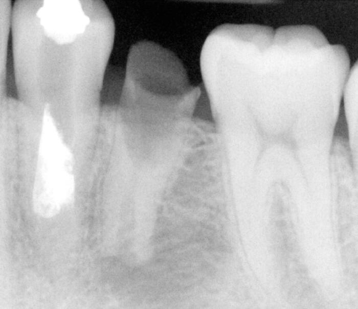
In fact, the tooth #2 is implant supported (Fig.2). Let us go back to Fig.1. You may notice the gum tissue around the tooth # 2 is as healthy as those around any teeth in the photo. The shade of the tooth #2 matches that of teeth above it. In contrast, natural teeth #1 and 3 look awkward. The teeth #1 and 2 originally suffer from a special form of congenital dental defect (Fig.3). This defect is fairly common found in Chinese children. The nerve of these two teeth get exposed, infected, and dead. The original tooth #2 cannot be saved due to failure of treatment and ensuring cavity. The tooth #1 received incomplete, inappropriate root canal treatment before. Dead nerve and incomplete root canal treatment contribute to the fact that the tooth #1 look so dark. We redo root canal treatment for the tooth #1 before placing an implant for #2. Root canal treatment is a type of preparation work we should do to eradicate infection. Fig.1 is taken approximately 6 months after implant. This young gentleman is now pursuing for doctor degree in the north. You may return to the main page of this lecture.
Xin Wei, DDS, PhD, MS 1st edition 02/14/2009, last revision 09/28/2012