|
|
|
|
|
|
|
|
|
Early Implantation
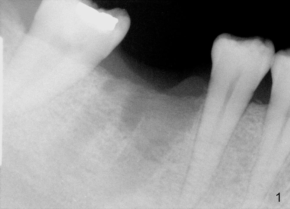
As compared to immediate implantation, early one occurs 4-8 weeks after extraction. The socket is initially healing with a lot osteogenic activities (producing bone), while the existing bone plate (especially buccal) and overlying gingiva have yet to shrink.
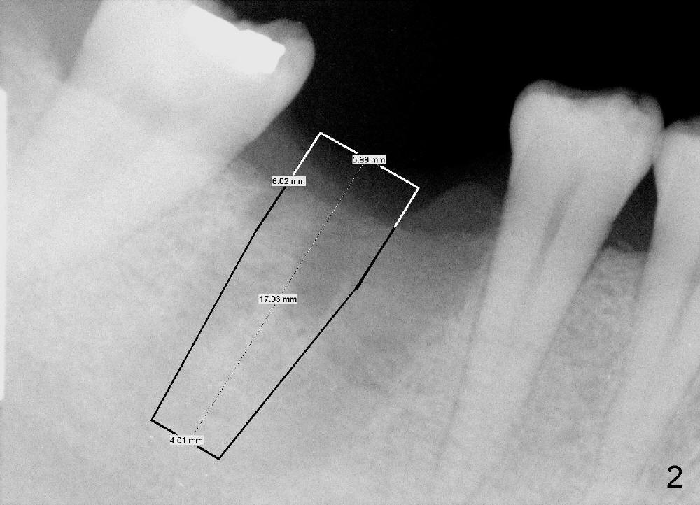
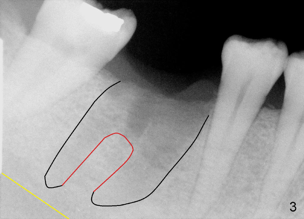
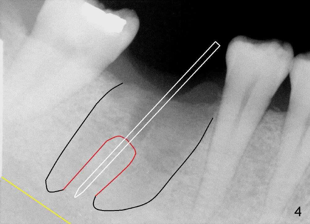
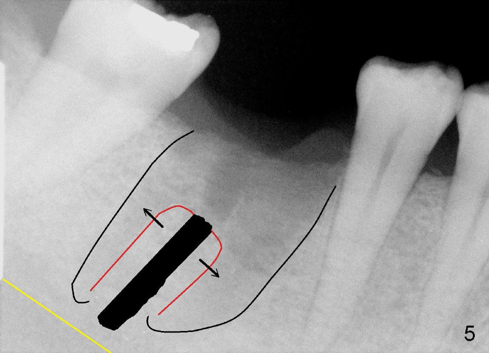
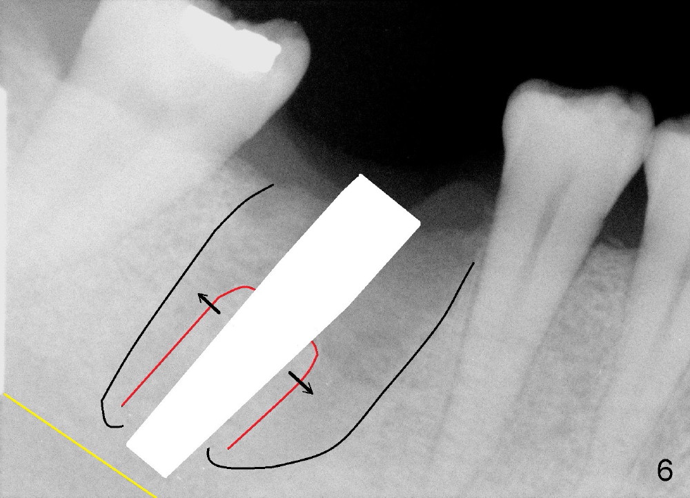
A 43-year-old lady had the lower right first molar extracted in other office last month. The socket appears to be healing normally (Fig.1). Tatum tapered implant 6x17 mm is planned (Fig.2). The implant is to be placed inside the septum (Fig.3 red outline; black: mesial and distal sockets; yellow: the upper border of the inferior alveolar canal). Either 1.5 mm pilot drill or 2 mm RT is used to start osteotomy in the septum at the depth of 17 mm (Fig.4 white outline). The osteotomy is enlarged with alternating use of osteotomes and 17 mm series tapered drills or Bicon reamers if autogenous bone will be needed (Fig.5 black), while the septal bone is pushed mesiodistally (arrows). After application of taps, an appropriately sized implant is placed (Fig.6 white). The septal bone continues expanding (arrows), whereas the mesial and distal sockets are being decreased.
The axis of osteotomy and of the implant is not consistent with that of the 2nd premolar. Instead it is more or less parallel to that of the 2nd molar. Intraop PA may confirm this observation.
Since the open of the socket has been sealed, there is no room for bone graft. But bringing bone graft materials to the operatory is not too redundant.
How is the surgery?
Xin Wei, DDS, PhD, MS 1st edition 09/22/2013, last revision 07/26/2014