|
|
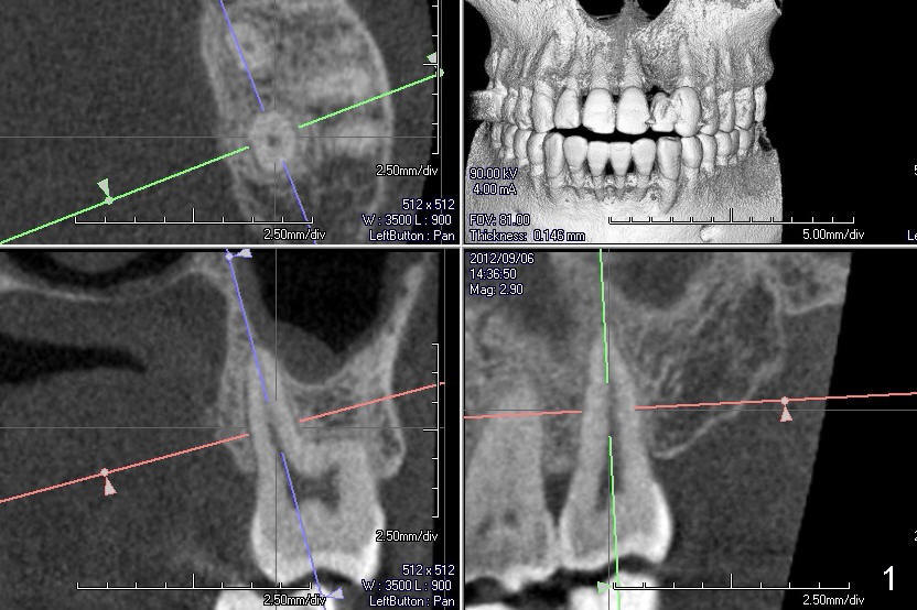
Fig.1: CT images from a different patient are used instead. The upper left image is an axial section, lower left coronal and lower right sagittal.
Return to Palatal socket or septum
Xin Wei, DDS, PhD, MS 1st edition 12/22/2013, last revision 12/22/2013