|
|
|
|
|
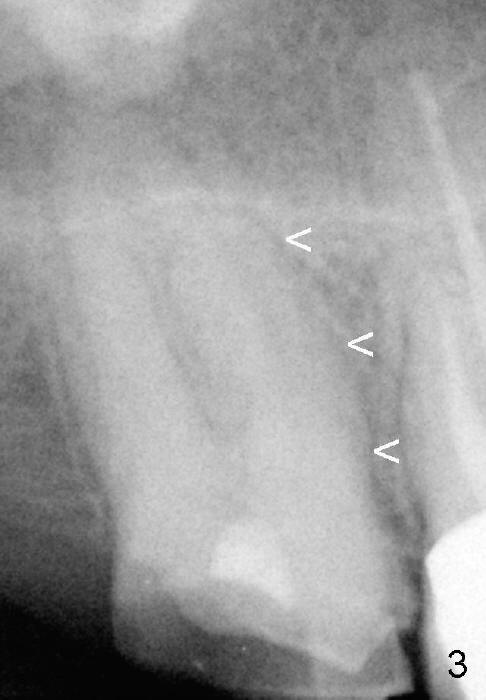
Fig.3 (preop PA) <: mesial lamina dura of the mesiobuccal root of the upper right 2nd molar.
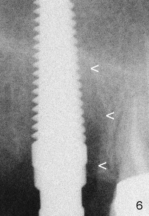
Fig.6 (intraop PA with 5 mm tap) <: mesial lamina dura of the mesiobuccal socket.
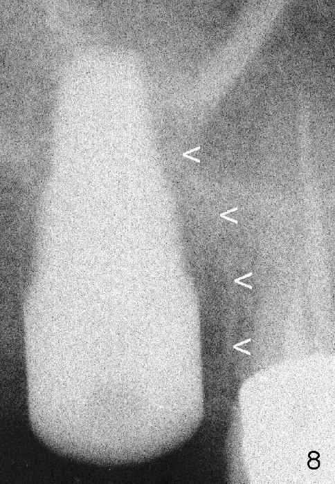
Fig.8 (immediately postop, 7x17 mm implant) <: mesial lamina dura of the shrinking mesiobuccal socket.
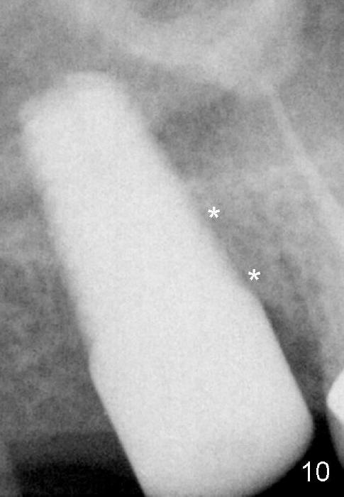
Fig.10 (5.5 months postop) * mesial socket space disappears (filled by new bone).
Xin Wei, DDS, PhD, MS 1st edition 05/05/2014, last revision 09/07/2014