|
|
|
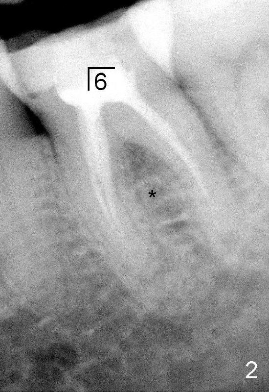
Root canal filling of the lower left 1st molar is incomplete with mild periapical radiolucency (Fig.2). The septum is wide (*).
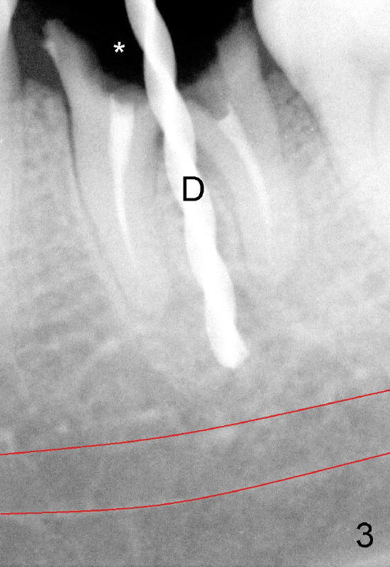
After removal of the remaining crown (Fig.3 *) and exposure of the top of the septum, a pilot drill (1.5 mm) is used to initiate the osteotomy (D). Red line: the upper and lower borders of the inferior alveolar canal.
Return to Immediate Implant in the Septum
Xin Wei, DDS, PhD, MS 1st edition 03/24/2013, last revision 01/29/2014