|
|
|
|
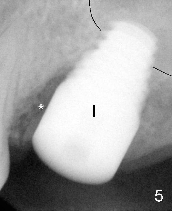
Fig.5 (immediately post implantation (I), 8x14 mm). *: remaining mesial socket
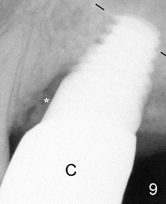
Fig.9 (5.5 months post implantation, immediately post cementation of the crown (C). The mesial socket has apparently decreased in size and increased in density.
Black line: sinus floor.
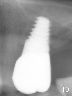
Fig.10: 34 months post cementation with closure of the mesial gap.
Return to Osteoporosis
Xin Wei, DDS, PhD, MS 1st edition 05/31/2013, last revision 10/04/2016