
|
|
|
|
|
|
|
|
|
|
|
|
|
|
|
|
|
|
||
"Instant Ortho"
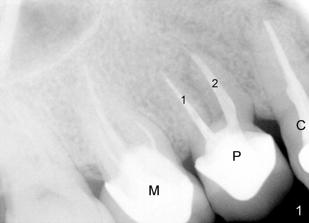
A 45-year-old man has multiple restorations. There is persistent pain in the upper right premolar (Fig.1 P, between canine (C) and the 1st molar (M)). This preop PA is taken several years before symptoms develop. It shows that this premolar is malpositioned: two roots (1,2) are aligned mesiodistally. The distal periodontal pockets are deep, consistent with root fracture.
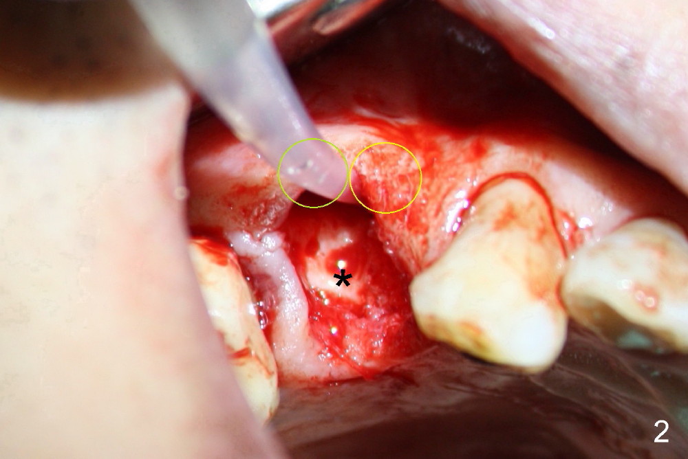
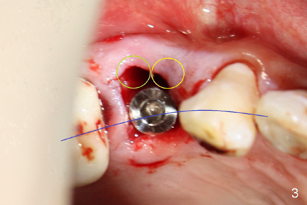
When the tooth is extracted, the two sockets (Fig.2 yellow circles, where the irrigation syringe is) are found to be buccally located, creating cantilever effect and probably leading to restoration failure/root fracture. There is bone palatal to the sockets (*). Bicon reamers are used to form osteotomy and collect autogenous bone at the same time (because of 50 RPM). A 5x17mm implant is placed, in alignment with arch form (Fig.3 blue curved line). The future restoration will be along the long axis of occlusion. Therefore the immediate implant achieves "instant orthodontic result".
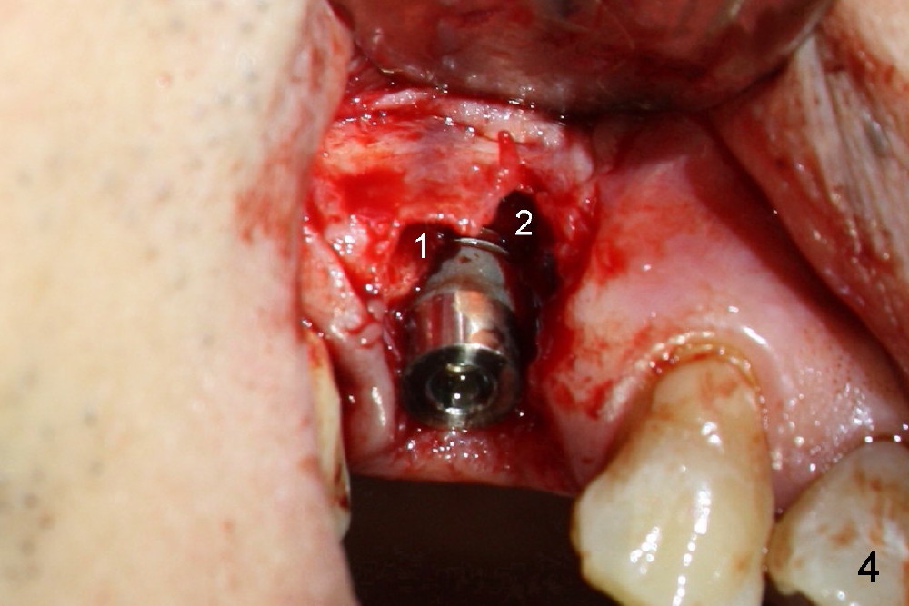
Buccal flap is raised (Fig.4) for bone graft and collagen membrane. The graft is autogenous, harvested using the reamers mentioned above and mixed with Bicon Synthograft.
The flap (F in Fig.5, taken 2.5 months postop) is closed following the periosteum being underscored.
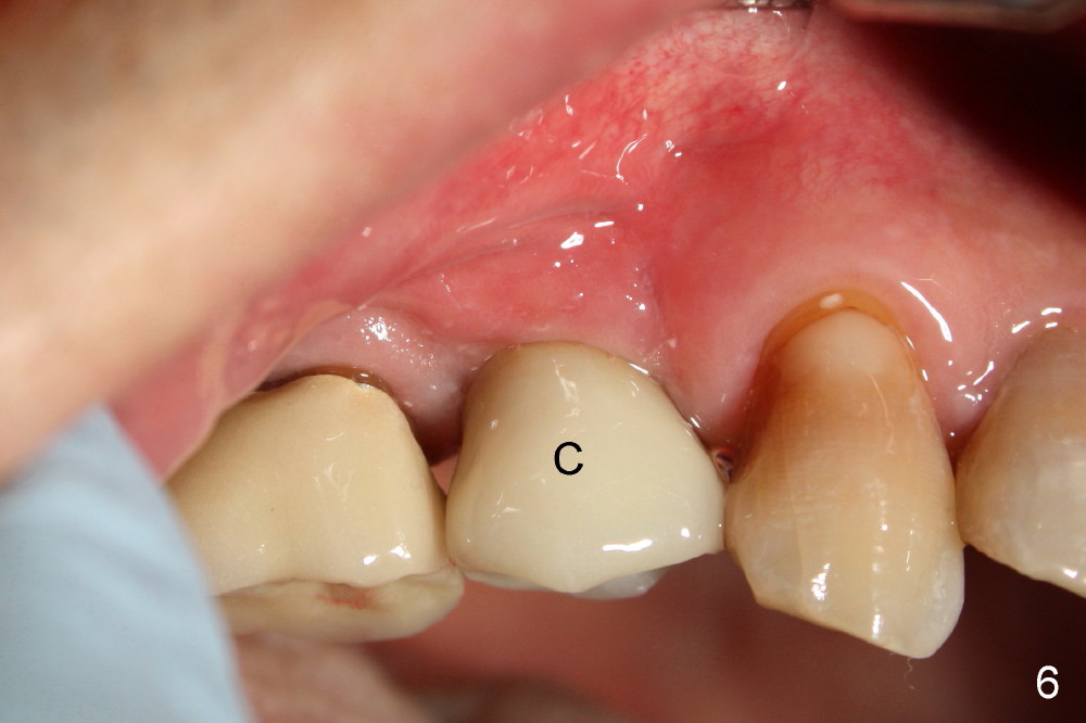
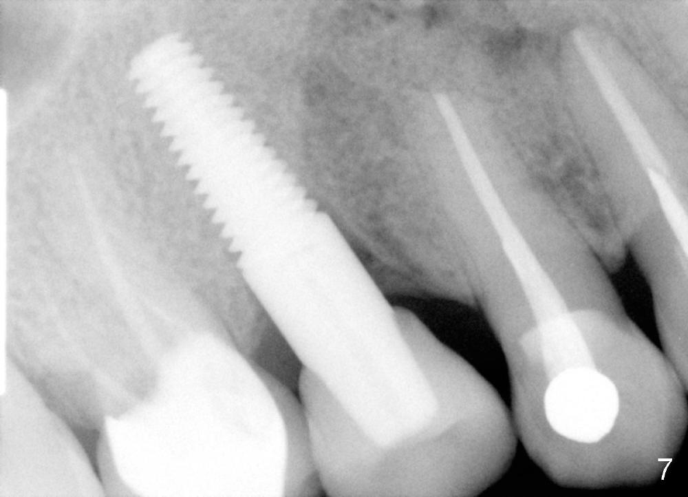
A single crown is cemented 3 months postop (Fig.6 C). Fig.7 is taken 6 months post cementation.
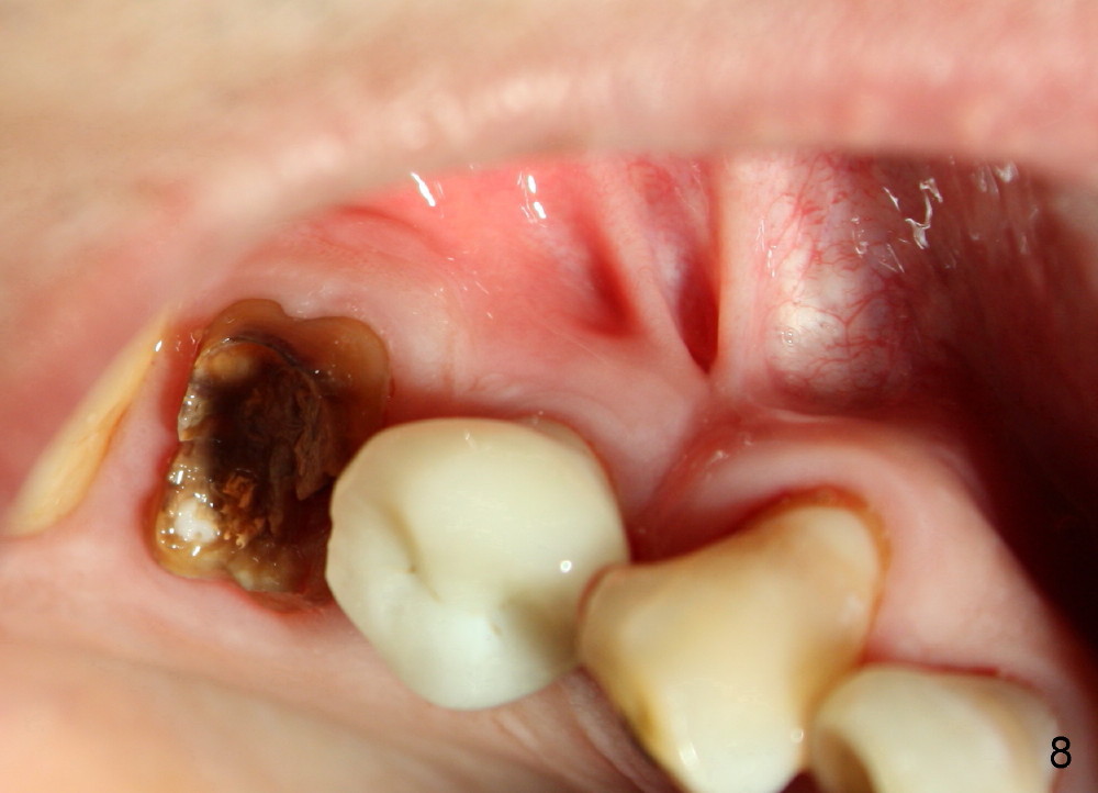
Eleven months post cementation, the patient returns for #3 crown recementation. The buccal plate atrophy over the implant is minimal (Fig.8).
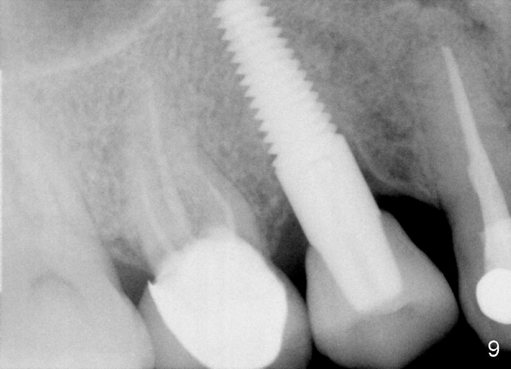
Fig.9 is taken 19 months post cementation of the implant crown. There is no bone resorption around the implant.
It appears that it is easier to place an immediate implant at the right position and angulation than delayed one. It is also easy to place an adjacent immediate one.
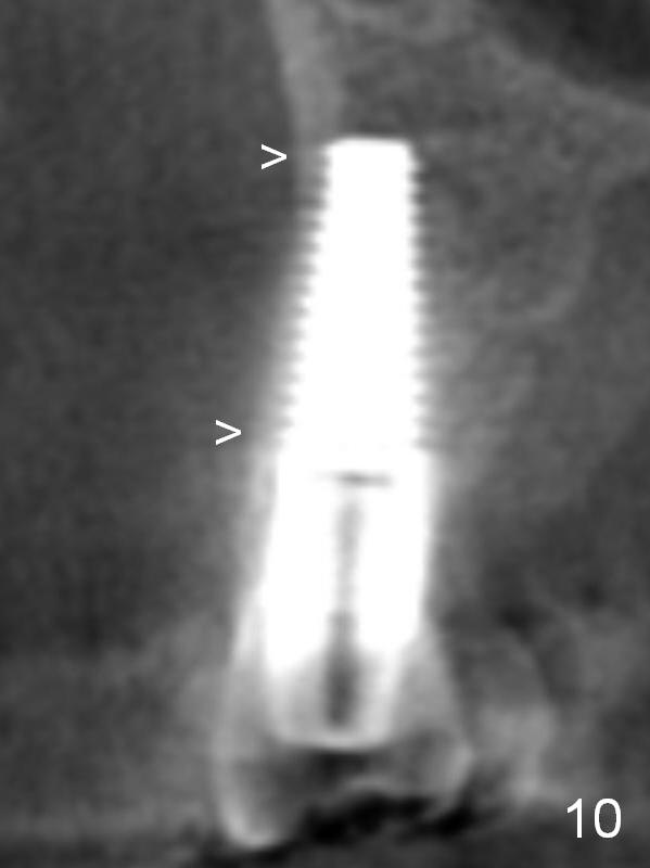
CT taken 3 years 1.5 months postop, 2 years 10 months post cementation shows the presence of the buccal plate, although thin (Fig.10 >, consistent with Fig.4).
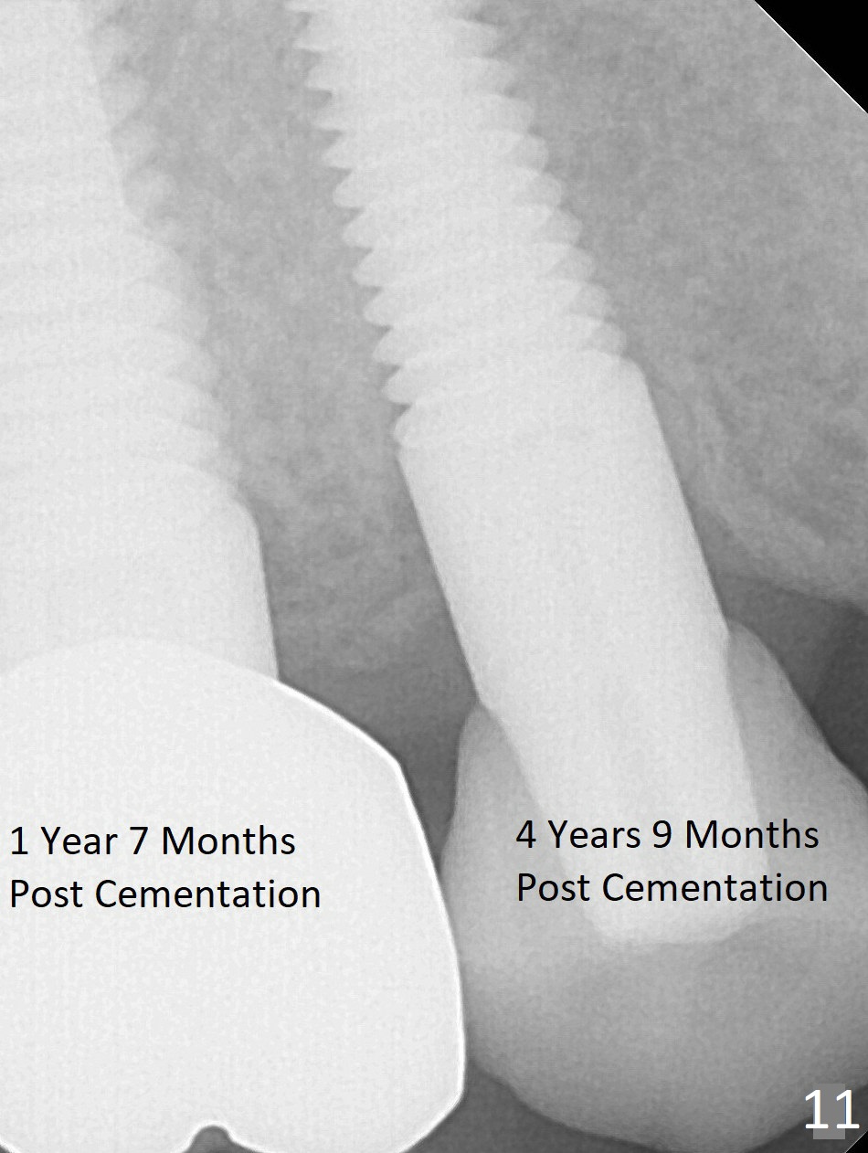
There is lamina dura-like structure mesially 4 years 9 months post cementation (Fig.11).
Return to Fellowship Documentation Form, Upper Bicuspid Immediate Implant, CT F-U
Xin Wei, DDS, PhD, MS 1st edition 03/30/2013, last revision 12/31/2017