|
|
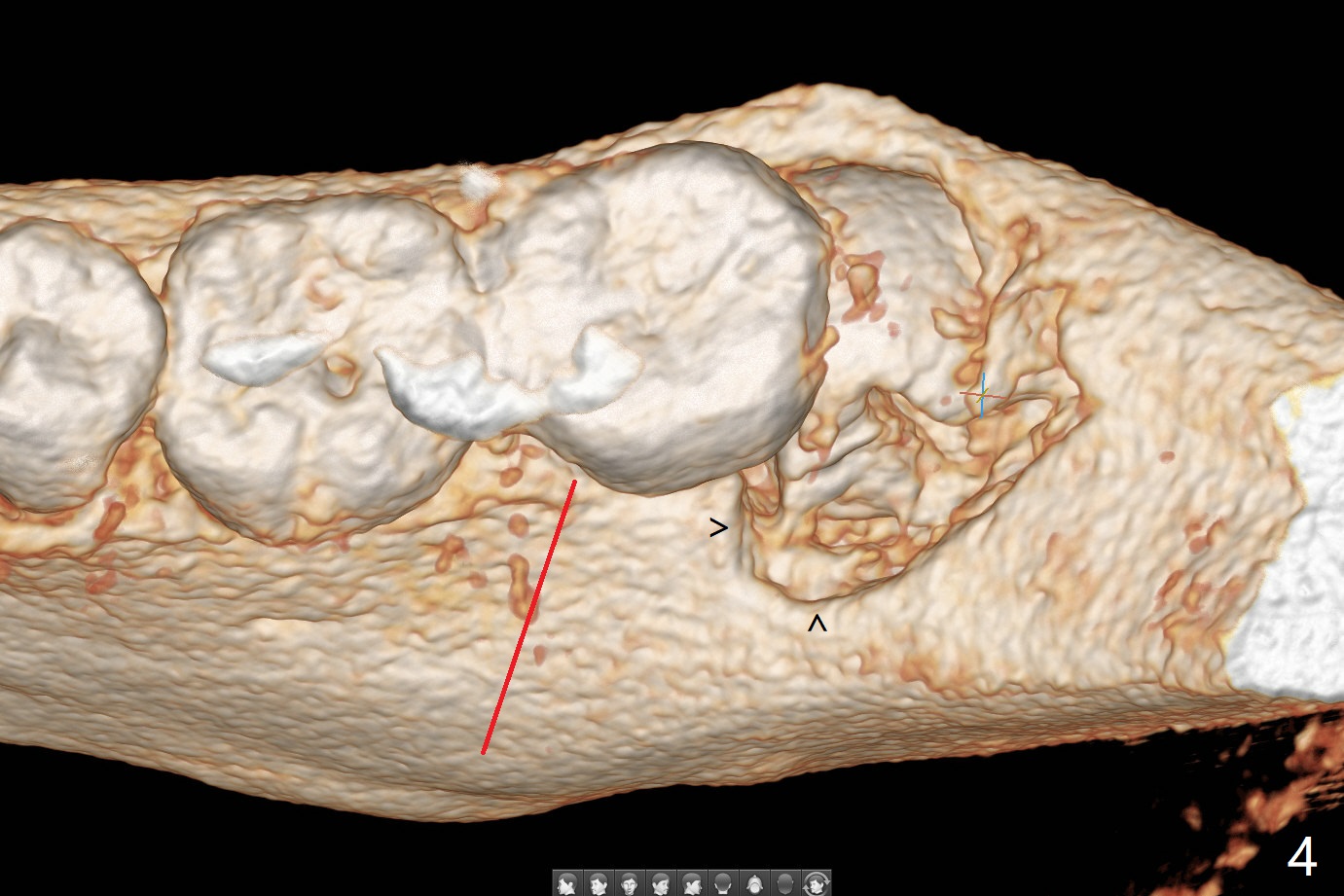
The accessory incision is placed mesiobuccal of the tooth #18 (Fig.4 red oblique line, Fig.6) so that it is not overlying the bony defect (Fig.3 arrowheads). Since the access to the impacted tooth is limited, small field of CT is taken (Fig.3,4), which shows the root is yet to be exposed (Fig.3 R). 内,外斜脊 (27岁女)
Grafts for 3rd Molars Last Next
Xin Wei, DDS, PhD, MS 1st edition 12/31/2019, last revision 07/31/2021