|
|
|
|
|
|
|
|
|
|
|
Augma vs. Osteogen Plug M
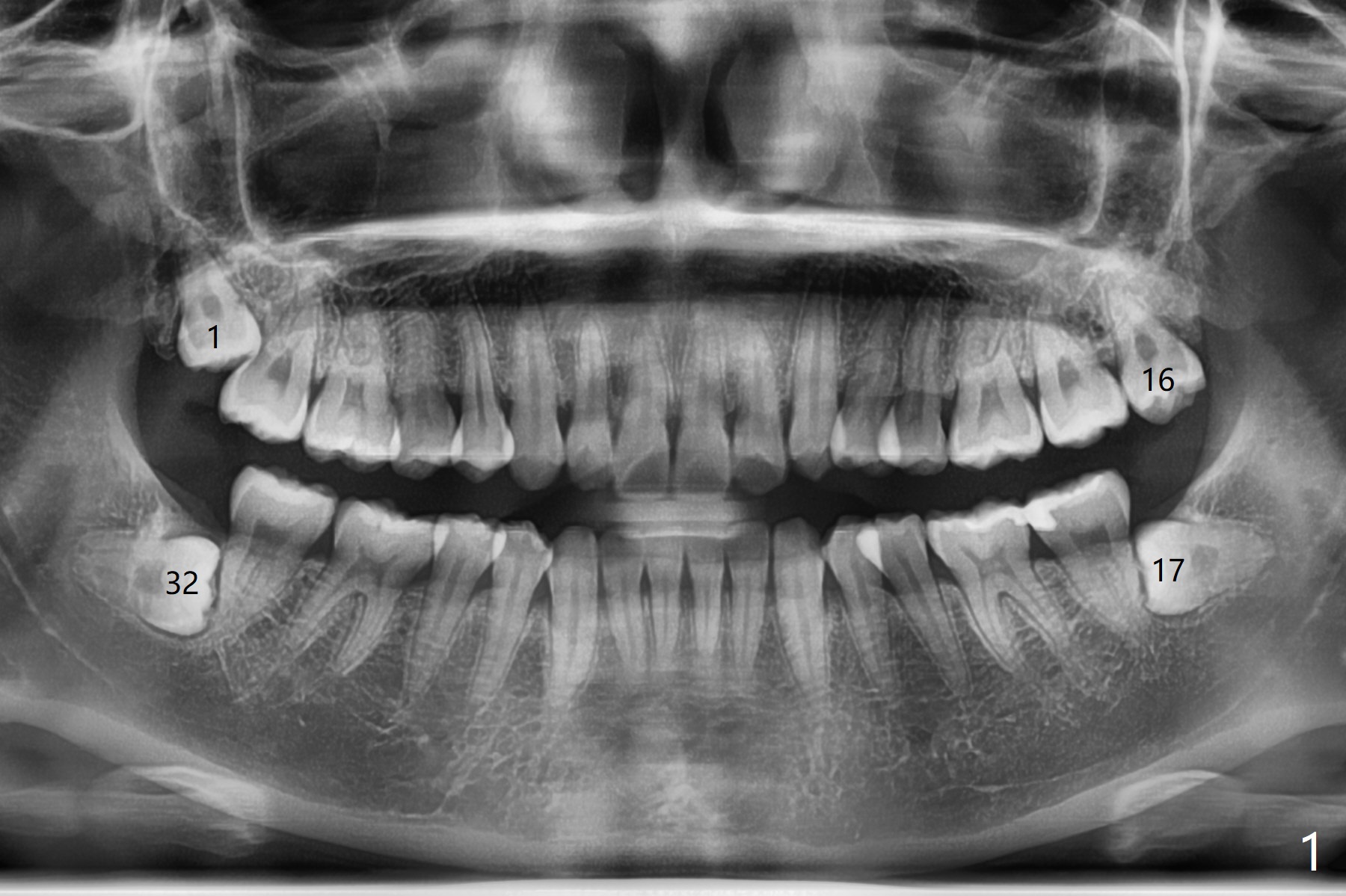
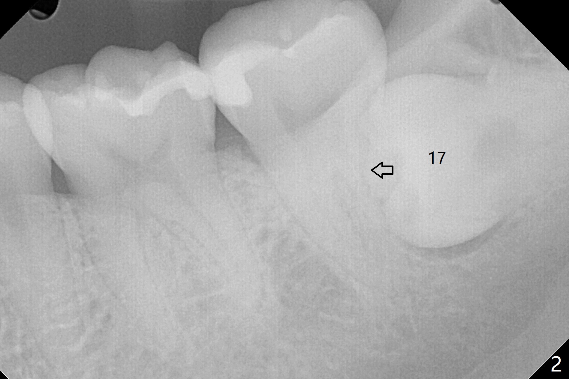
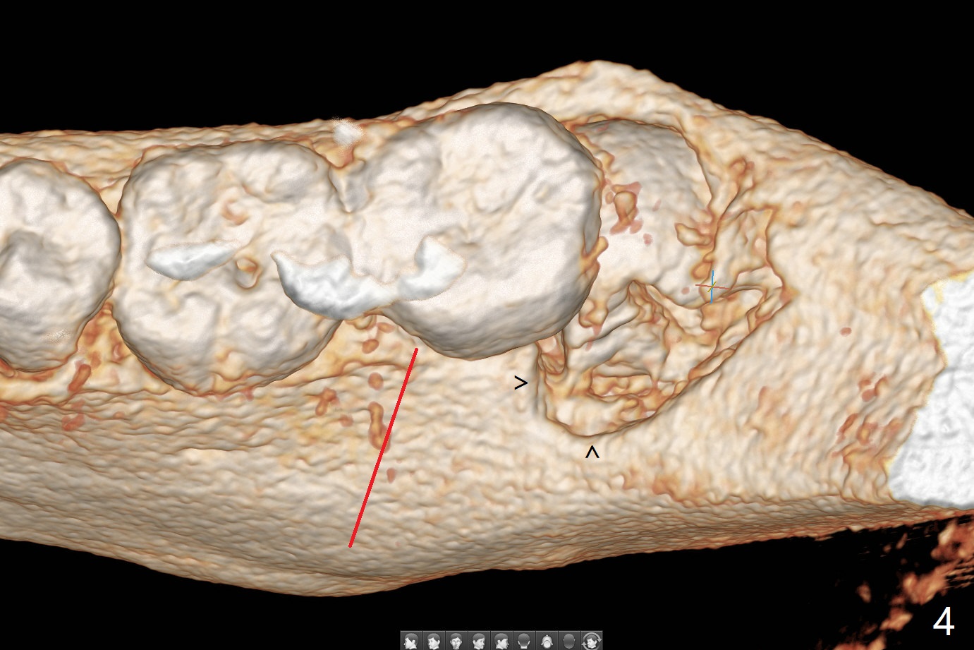
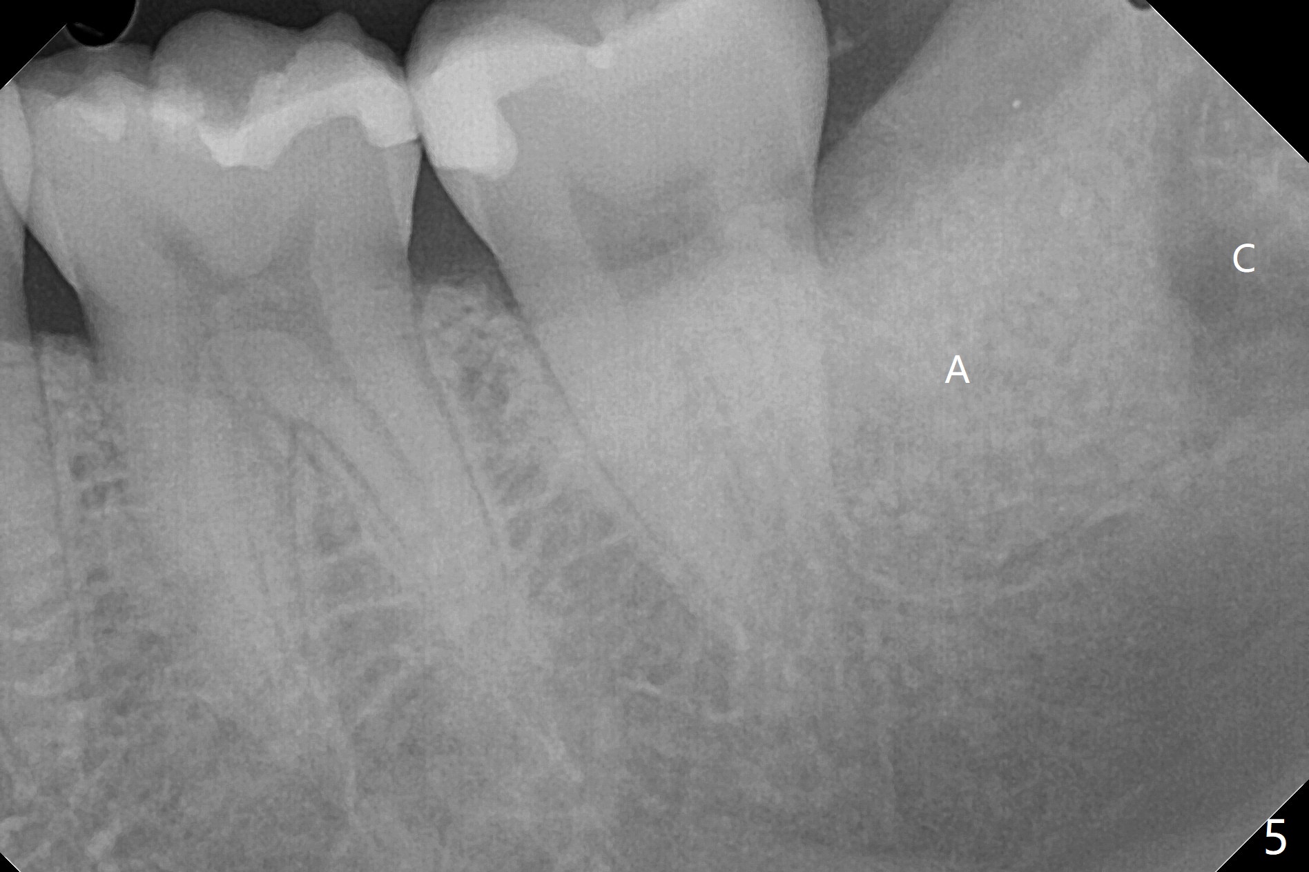
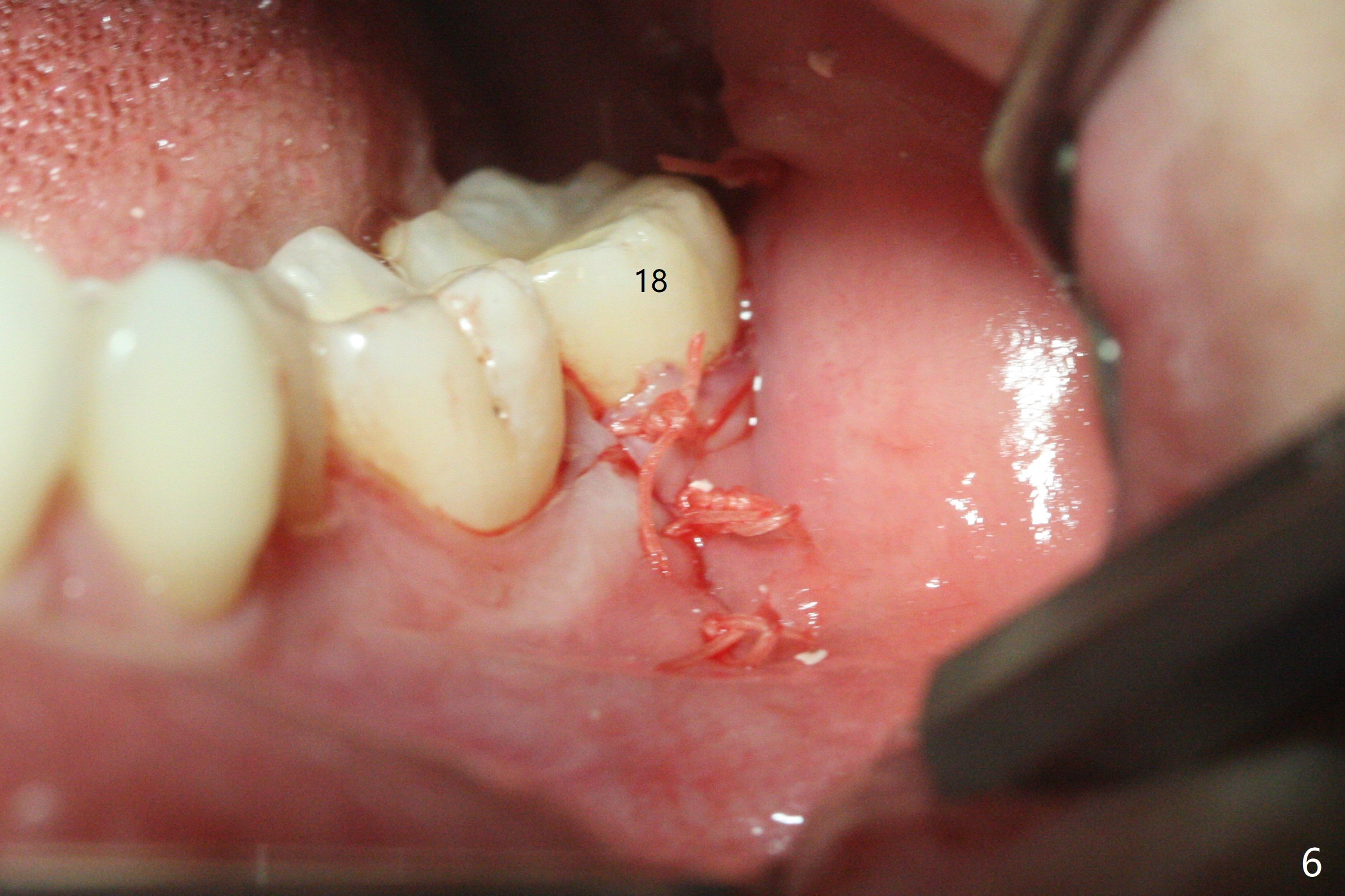
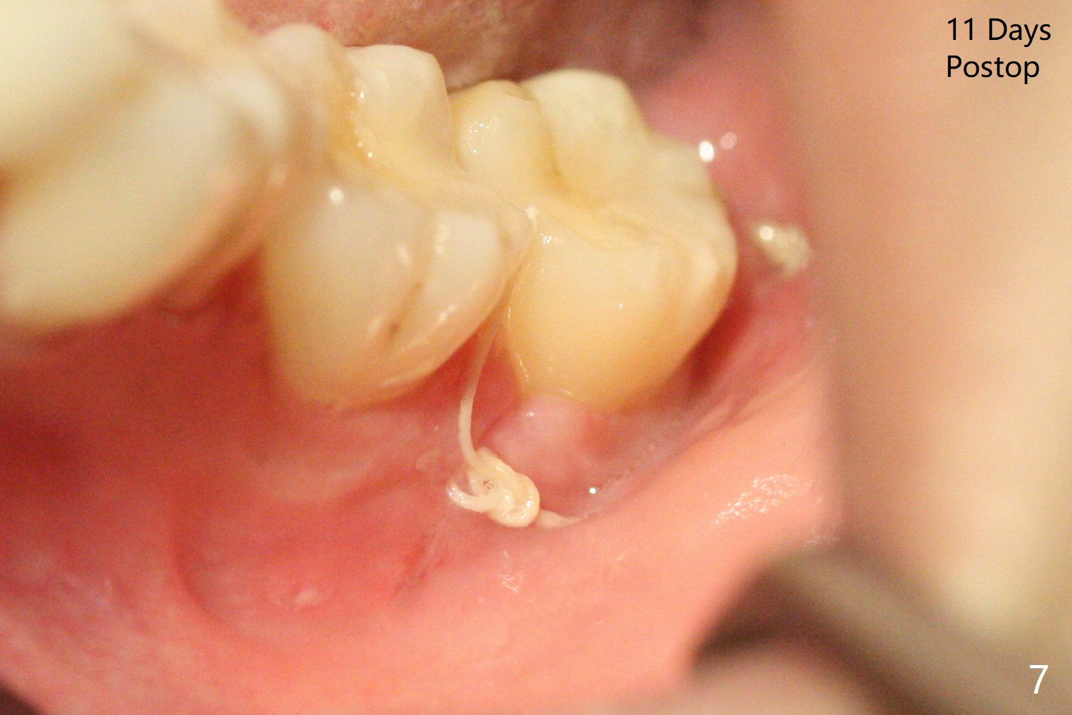
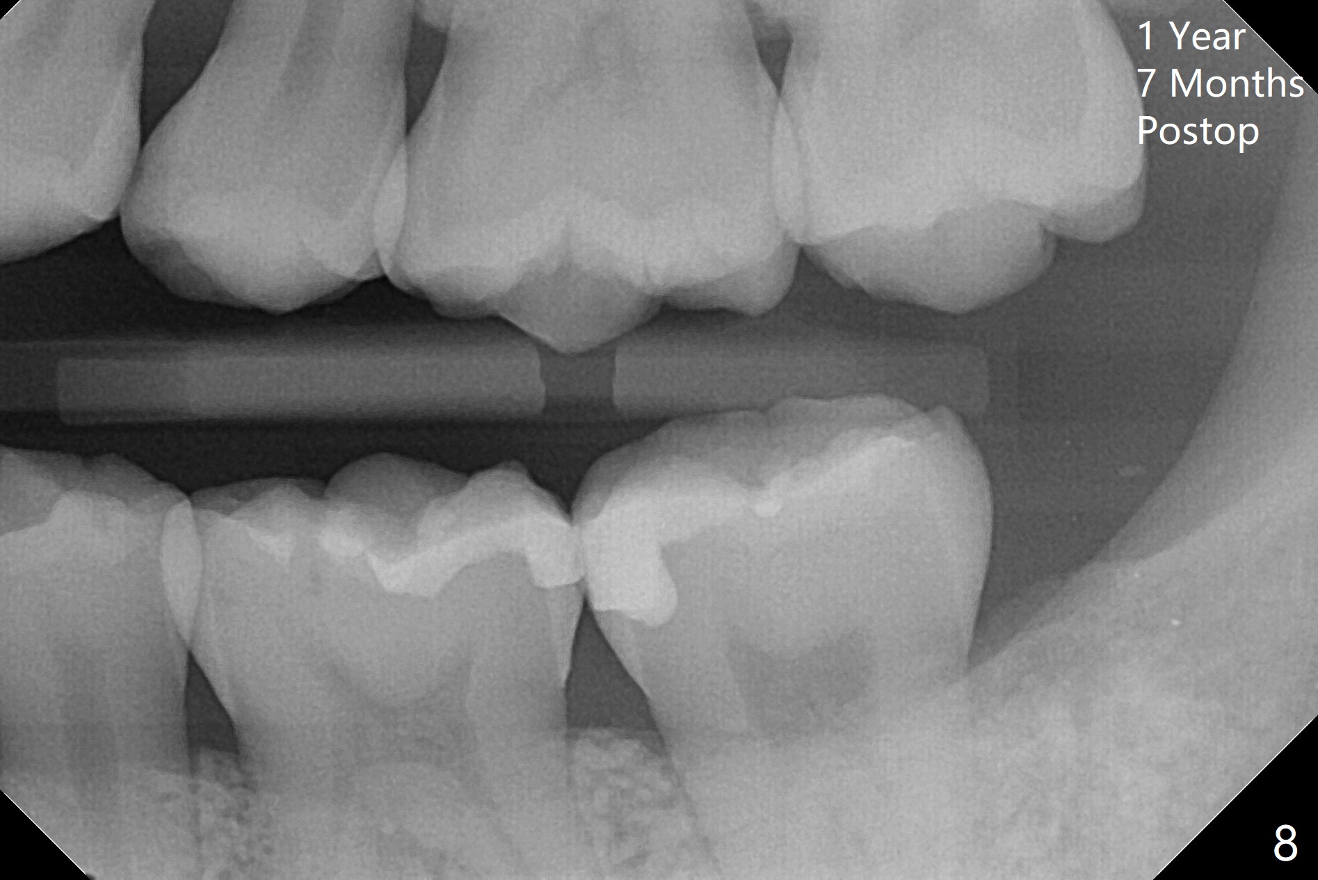
A 27-year-old woman (nervous) is going to return for #16 and 17 extraction (Fig.1). Offer sedative (Valium) if she cannot overcome fear. Take PAs for #17 and 32 to confirm Buccal Impaction, which dictates position of the accessory incision to reduce loss of bone graft in case of wound dehiscence. Place Collagen Plug (1/2 piece) in the apical portion of the sockets of the lower 3rd molars, while Augma and Osteogen Plug (1 piece) in the coronal half of #17 and 32, respectively. Place additional Collagen Plug for the remaining socket if needed before 4-0 PGA suturing as the 2nd step to decrease the chance of losing bone cement. Preop PA shows that the tooth #17 seems to be mesial (Fig.2 arrow). The accessory incision is placed mesiobuccal of the tooth #18 (Fig.4 red oblique line, Fig.6) so that it is not overlying the bony defect (Fig.4 arrowheads). Since the access to the impacted tooth is limited, small field of CT is taken (Fig.3,4), which shows the root is yet to be exposed (Fig.3 R). After tooth removal, Collagen plug is placed in the apex of the socket for hemostasis (Fig.5 C), while Bond Apatite coronal for bone regrowth (A). There is no dehiscence 11 days postop (Fig.7), although the patient complains of pain in the jaw and the temporomandibular region. The anterior portion of the external oblique ridge forms 1 year 7 months postop (Fig.8).
Return to Plug Augma 智齿拔除 Xin Wei, DDS, PhD, MS 1st edition 12/27/2019, last revision 07/25/2021