|
|
|
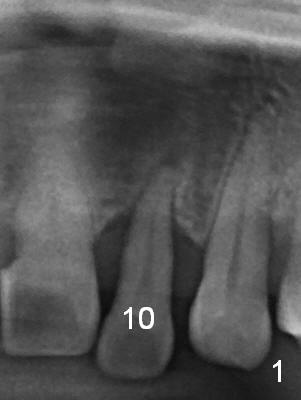
Prior to the surgery, the patient mentions that the future tooth at the site of #10 should not imitate the one in the drifted position (Fig.1 trimmed from panoramic X-ray).
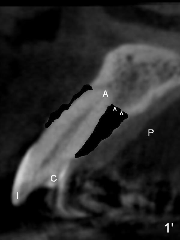
Fig.1' is CBCT cross section of the tooth #10 from another patient with illustration to show that the palatal (P) bone is flat (arrowheads) before extraction.
A: apical socket; C: cingulum; I: incisal edge.
Xin Wei, DDS, PhD, MS 1st edition 05/29/2015, last revision 05/30/2015