|
|
|
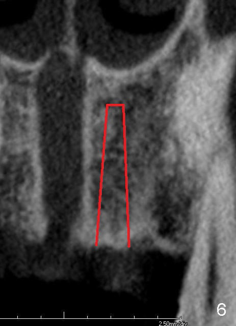
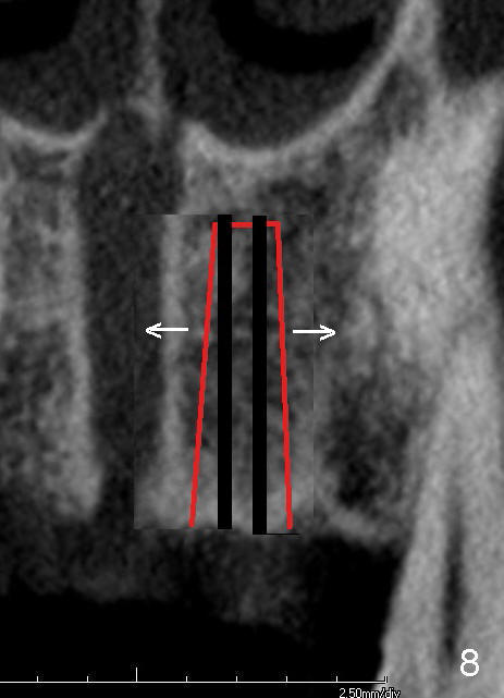
The osteotomies are under prepared (Fig.6 (from CT coronal section) red lines) and then expanded with taps and implants, pushing the wall of the nasopalatine canal medially (Fig.8 arrow).
Return to Incisive Canal
Xin Wei, DDS, PhD, MS 1st edition 06/02/2013, last revision 01/19/2018