|
|
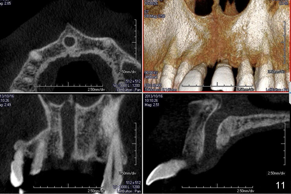
Fig.11: After surgery, the nasopalatine canal is studied using preop CBCT scans (axial (upper left), coronal (lower left), and sagittal (lower right) sections and 3 D image (upper right)). The canal is enlarged and deviated to the left.
Return to Incisive Canal
Xin Wei, DDS, PhD, MS 1st edition 06/02/2013, last revision 01/19/2018