


|
|
|
|
|
|
|
|
|
|
|
|
|
|
|
|
||
Extra Precaution Measure
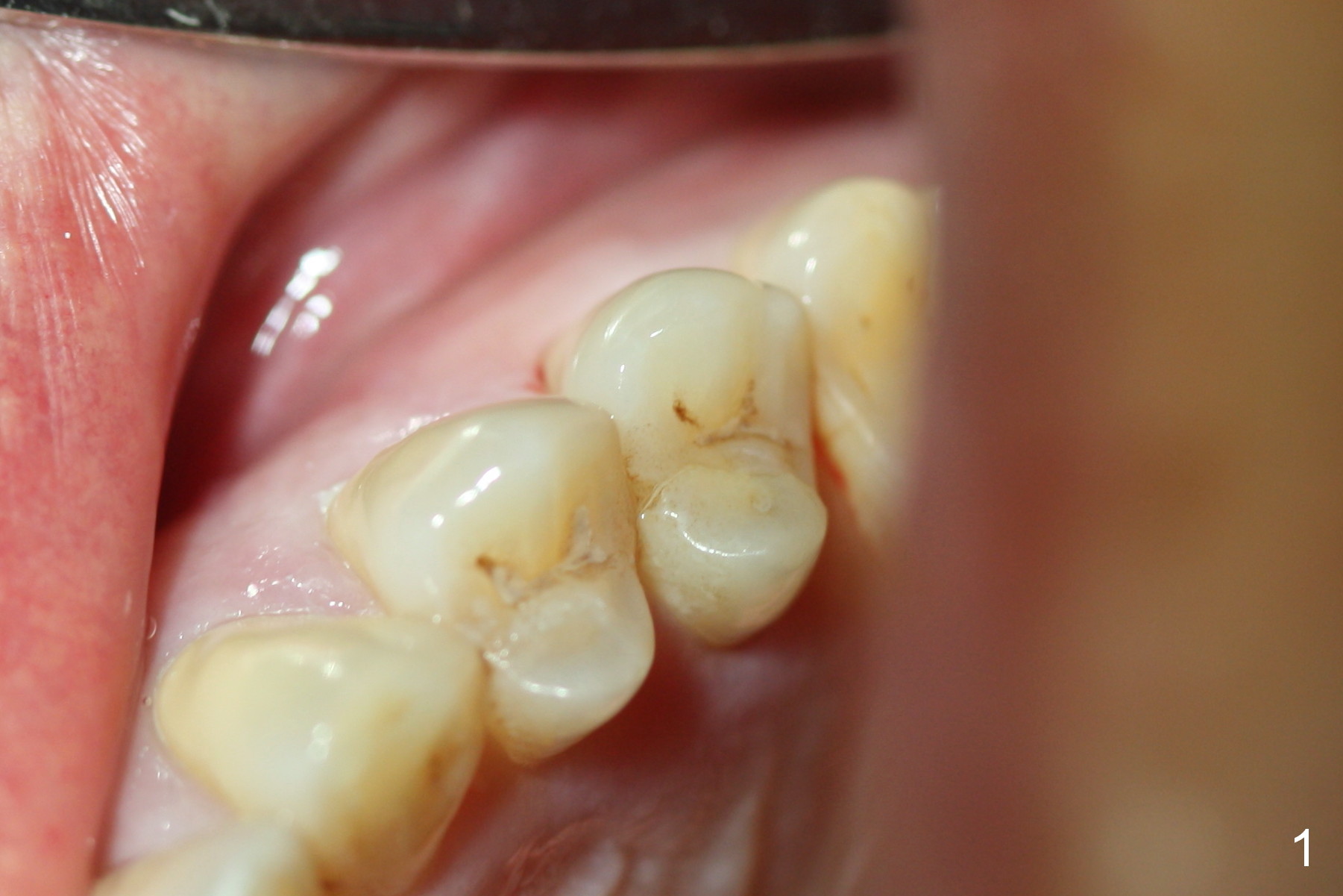
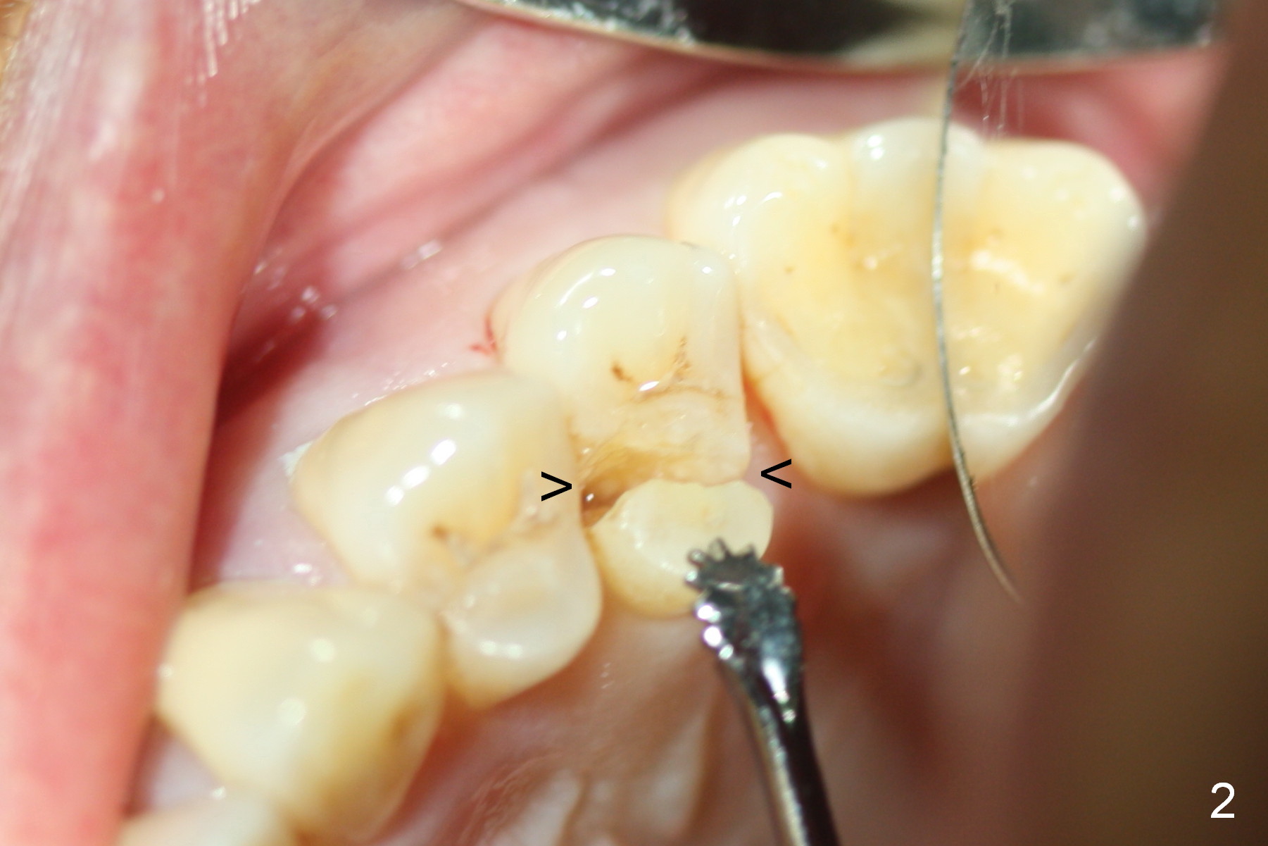
Fig.1,2 show preop condition. Extraction turns out to be not so difficult as expected. There is bone resorption in the palatal crest, as related to the fracture. The bone density does not feel high during osteotomy. So a long implant is planned. The depth of osteotomy is 20 mm (Fig.3,4 with 2.5 mm reamer). The trajectory is off; the red dashed line represents the socket. The trajectory is too late to change: when a 4.5x20 mm tap is inserted, the trajectory does not improve (Fig.5 T). The first intraop PA should have been taken when a 2 mm pilot drill was 14-17 mm deep. A 4.5x20 mm implant is placed with insertion torque at >56 Ncm (Fig.6 I). A 3.5x3 mm abutment is placed and prepared (A). Allograft is placed in the palatal crest prior to implantation. More bone graft is placed in the buccal gap after implantation, followed by Osteoplug (Fig.7 G). An immediate provisional is cemented to close the oval socket (Fig.8 P).
The patient reports mild hemorrhage a few hours postop. If extra amount of collagen membrane (dressing) were placed around the implant and on the top of Osteoplug prior to cementation of the provisional, the hemorrhage would have been less. This is the extra precaution measure to be taken.
The apprehensive patient is pleased when she returns for follow up 8 days postop. The buccal gap has closed (Fig.9 <, as compared to Fig.8). Although there is no tenderness, the palatal gingiva is slightly erythematous, corresponding to bone resoprtion in the palatal crest (related to the fracture line, Fig.2). It would be safer to place an implant slightly away from the palatal wall when the palatal fracture line extends subgingivally. Without proper communication with technician, the buccal margin of the definitive crown can be not ideal. There is no bone loss 20 months post cementation (Fig.11).
Return to
Upper Bicuspid Immediate Implant
Xin Wei, DDS, PhD, MS 1st edition 09/09/2015, last revision 01/19/2018