|
|
|
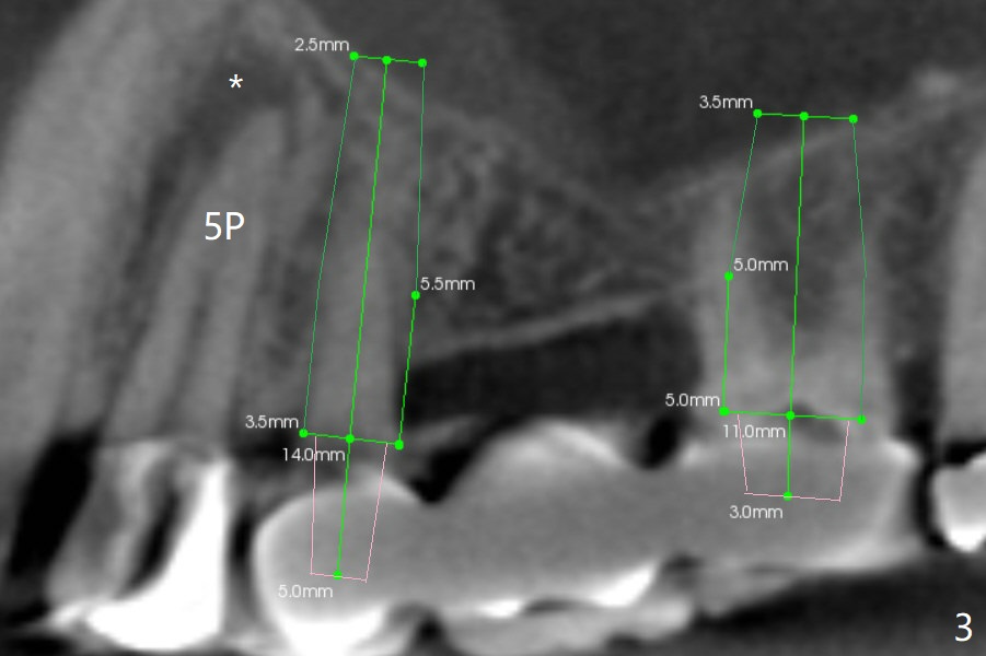
Furthermore CT more clearly shows apical lesions of the tooth #5 (Fig.3 (sagittal section)) than PA (Fig.2). In addition to pulpal test, RCT should be done for #5 prior to implant placement at #4. *: periapical radiolucency associated with the palatal root (P) of the tooth #5.
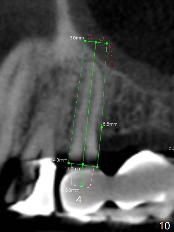
Fig.10 is a more precise sagittal section for #4. It appears that a 4x17 mm tissue-level implant is appropriate for the site.
Xin Wei, DDS, PhD, MS 1st edition 10/22/2017, last revision 01/19/2018