|
|
|
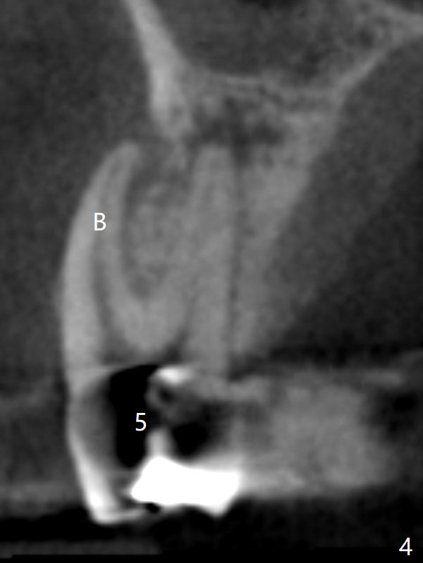
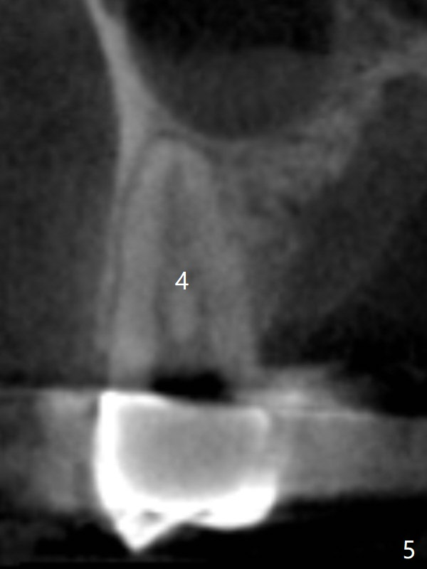
CT more clearly shows apical lesions of the tooth #5 (Fig.4 (coronal section)) than PA (Fig.2). Periapical radiolucency is associated with the palatal root of the tooth #5, while the buccal (B) root seems to be outside bone boundary. In addition to pulpal test, RCT should be done for #5 prior to implant placement at #4 (Fig.5). If an implant is needed at #5, it should be placed in the palatal socket with small diameter.
There is limited bone immediately apical to the apex of the tooth #4.
Xin Wei, DDS, PhD, MS 1st edition 10/22/2017, last revision 01/19/2018