|
|
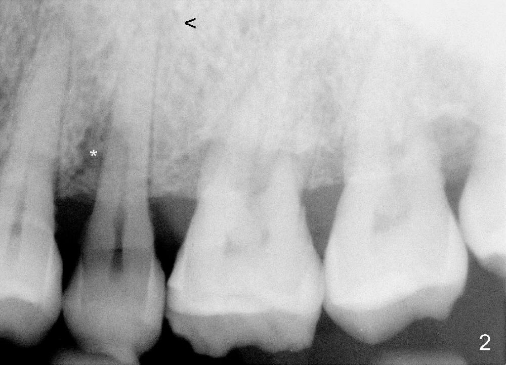
Preop PA shows periapical (Fig.2 <, due to pulpal exposure) and periradicular (*, due to subgingival fracture) radiolucency.
In spite of the fact that there is severe bone loss involving the 1st and 2nd molars, the PA does not show the upper portion of bone. The next PA is to be taken when the tooth is extracted to confirm that a long implant (20 mm long) is proper for the site.
Palatally Placed Tapered Implant Last Next
Xin Wei, DDS, PhD, MS 1st edition 12/17/2014, last revision 12/29/2018