|
|
|
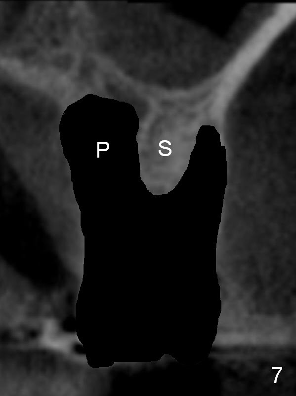
Fig.7 is an illustration of CBCT coronal section after extraction. There is severe bone resorption in the palatal socket (P). The septum is smaller and shorter than 22 months earlier.
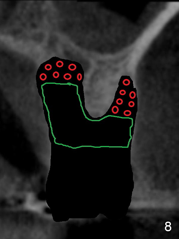
After debridement of the sockets, allograft mixed with Osteogen is placed in the deepest portion of the sockets (Fig.8 red circles), while the rest of sockets is filled with Osteogen plug (green outline).
Xin Wei, DDS, PhD, MS 1st edition 06/25/2015, last revision 06/25/2015