
,%20Vera.jpg)
|
|
|
|
|
|
|
|
|
|
|
|
|||
Buccal Placement
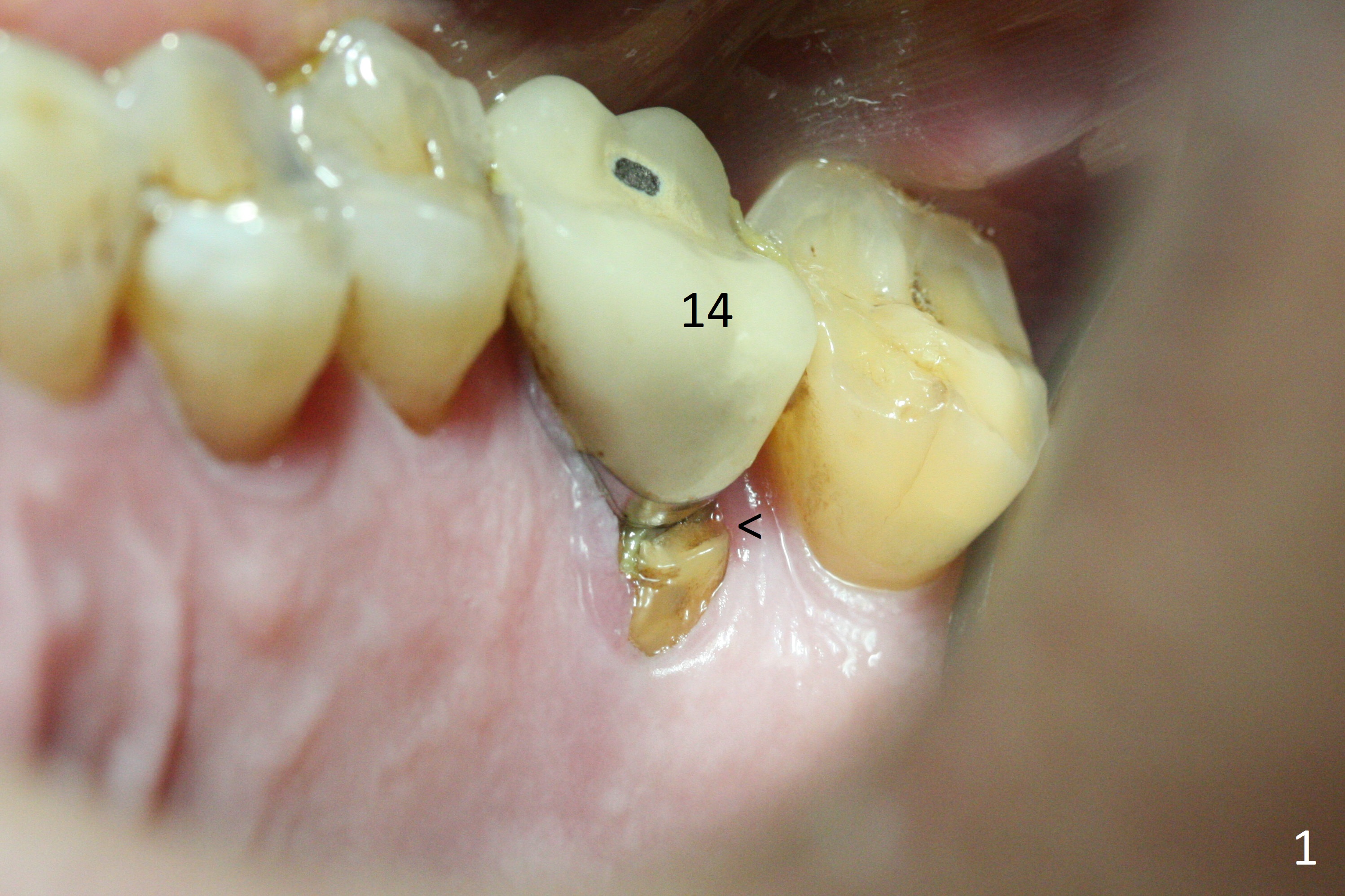
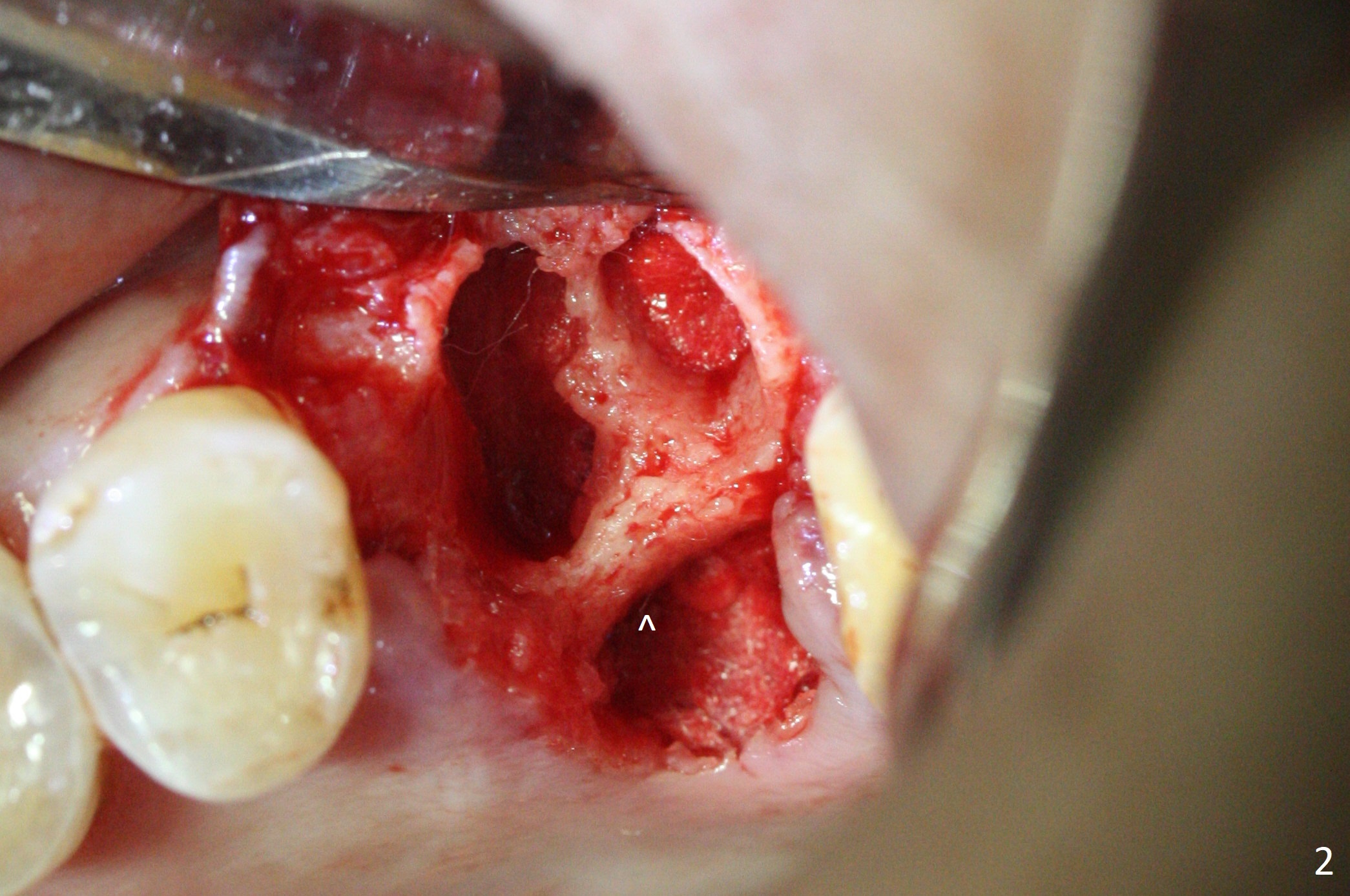
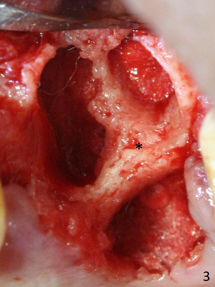
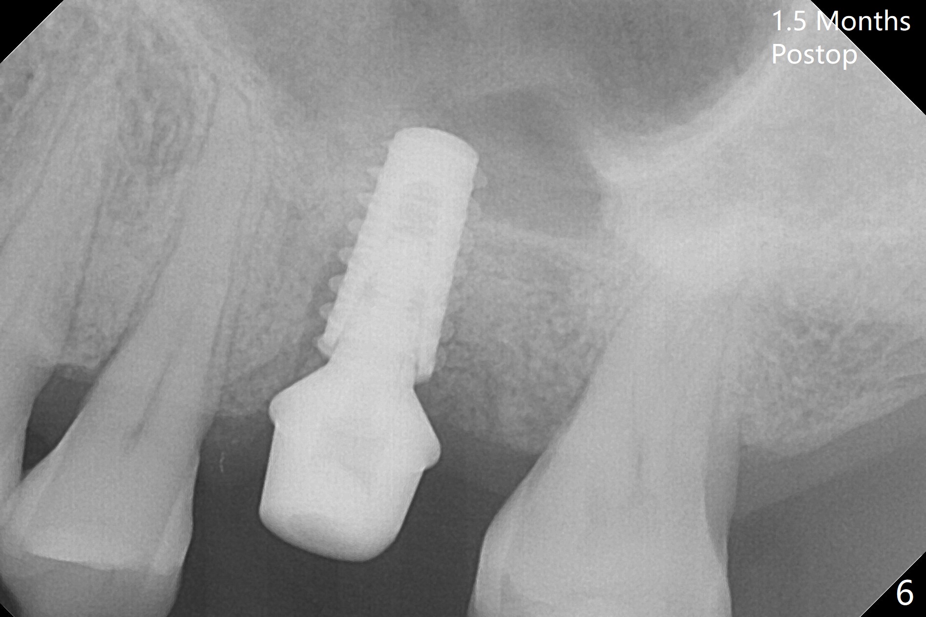
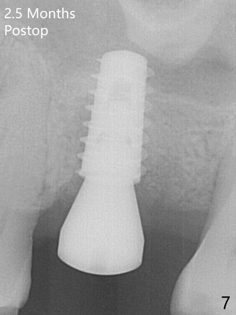
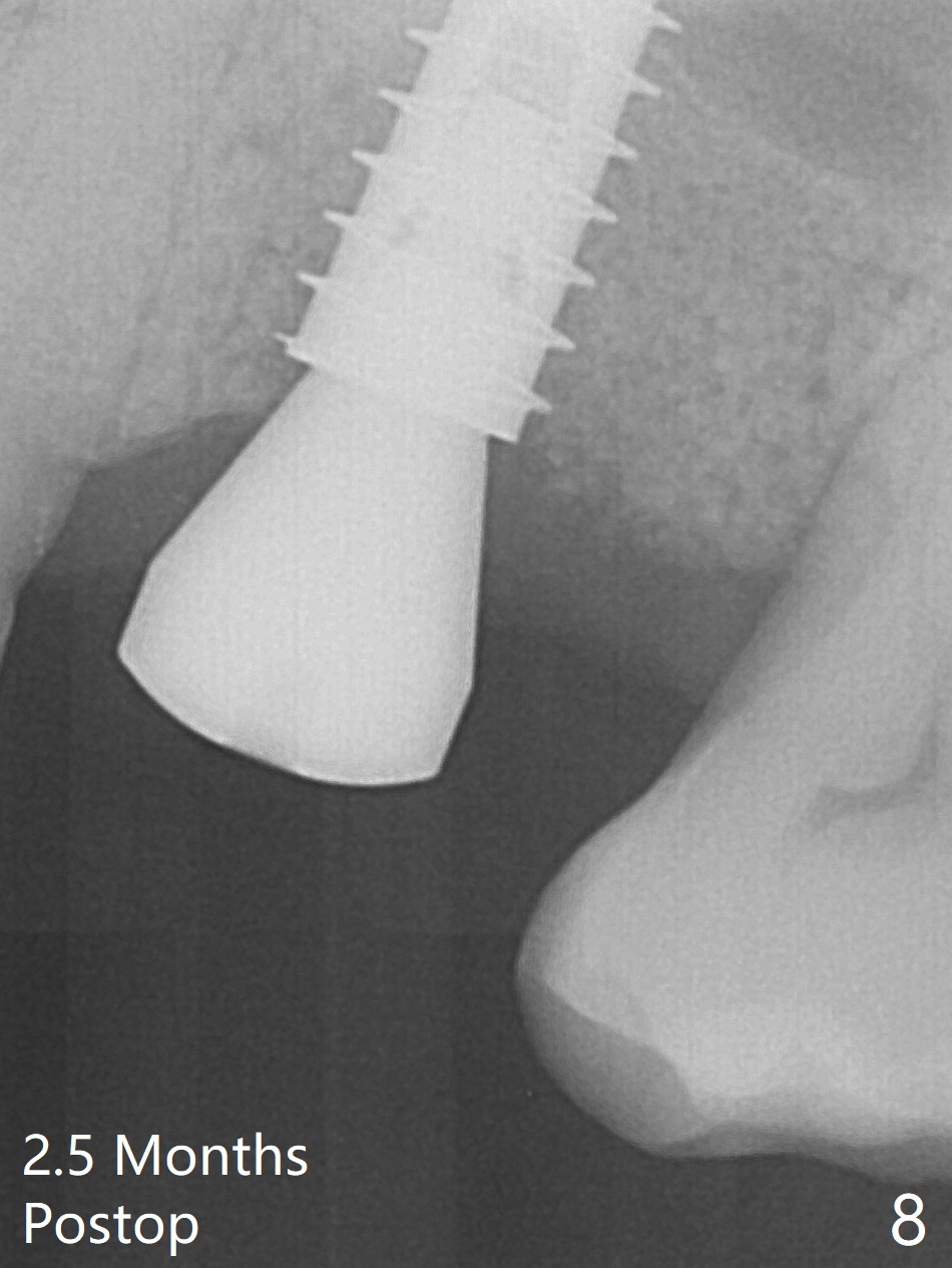
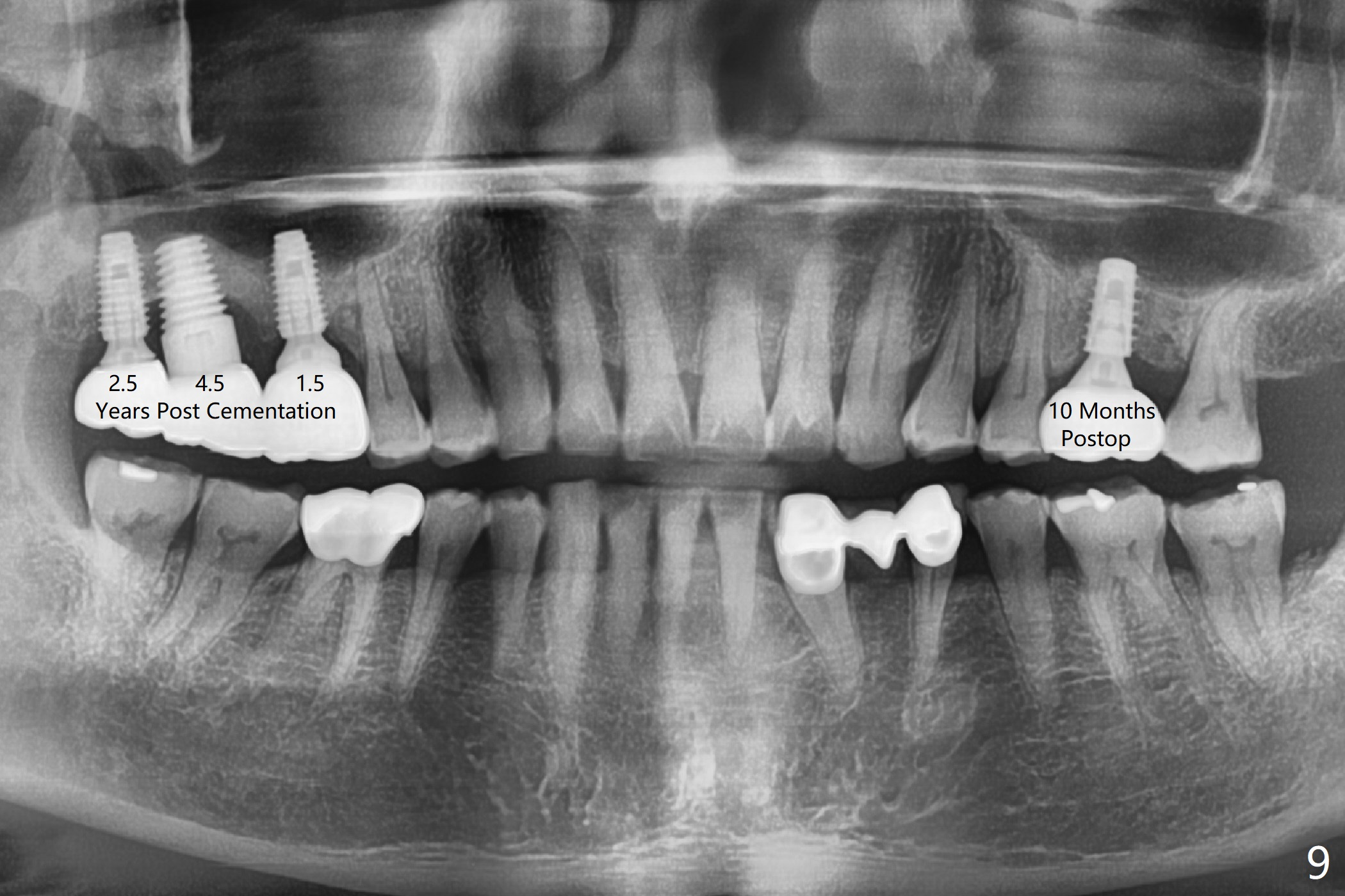
After extraction of the tooth #14 with palatal root fracture (Fig.1 <), sinus membrane perforation is found in the buccal wall of the palatal socket (Fig.2 ^). Osteotomy is initiated in the buccal strut of the septum (Fig.3 *). As the osteotomy increases in diameter, it slides into the mesiobuccal socket (Fig.4). A 5x9 mm implant is placed slightly mesial; after placement of a 6.5x4(2) mm abutment and insertion of collagen plug in the palatal socket, Vera graft is placed in the remaining socket space (Fig.5 *). Nasal hemorrhage persists 1.5 months postop (Fig.6). The distal gingiva is slightly tender and erythematous; a 6x4 mm healing abutment is placed 2.5 months postop (Fig.7,8). Impression is taken after laser gingivectomy 3.5 months postop. The patient feels discomfort at the site 7 days post impression, although the gingiva heals after laser treatment. Two weeks after cementation, the mesial gingiva is mildly tender and the abutment screw is loose. A 6x3 mm healing abutment is placed. The abutment/crown is reseated 10 months postop (Fig.9).
Return to
Upper
Molar Immediate Implant, Prevent
Molar Periimplantitis (Protocols,
Table),
Armaments
1
2
3
Xin Wei, DDS, PhD, MS 1st edition 12/11/2017, last revision 12/09/2018