|
|
|
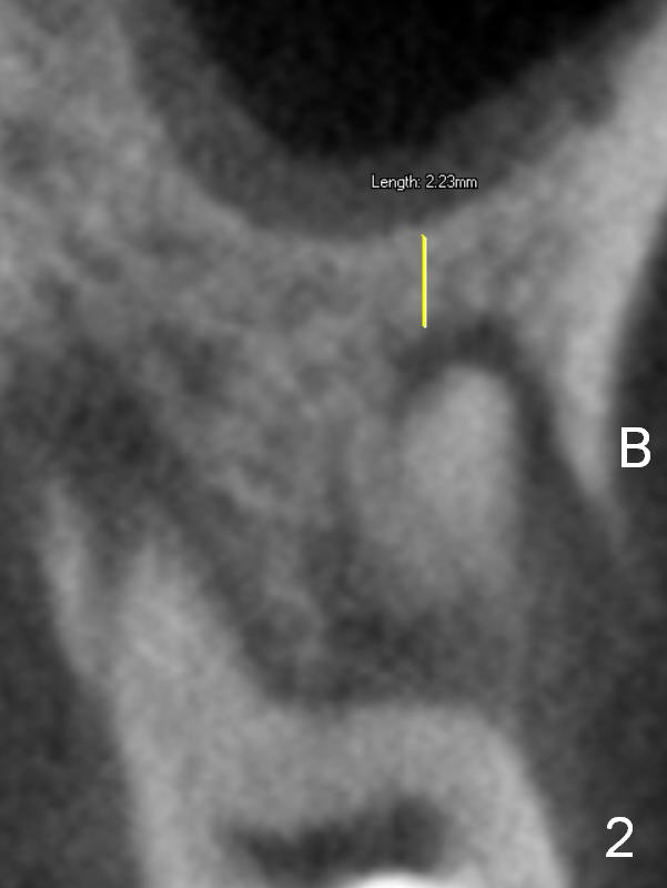
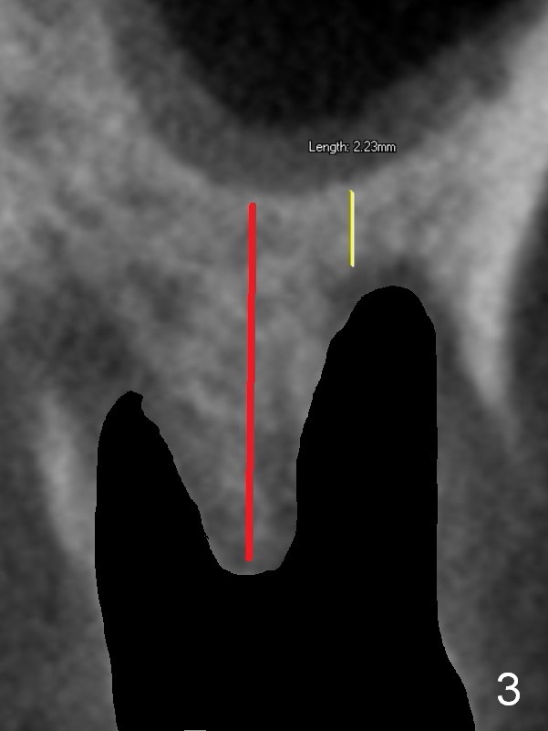
There are periapical radiolucent lesions of the buccal (Fig.2 (CT coronal section) B) and palatal roots; bone height above the buccal apex is ~ 2 mm (Fig.3). When the tooth #14 is extracted, a 1.6 mm pilot drill is used to start osteotomy in the middle of the fairly thin septum (Fig.3 red line, Fig.4 S).
Implant Placed in Thin Septum Last Next
Xin Wei, DDS, PhD, MS 1st edition 08/02/2016, last revision 06/03/2018