|
|
|
|
|
|
|
|
|
|
Exploratory Procedure
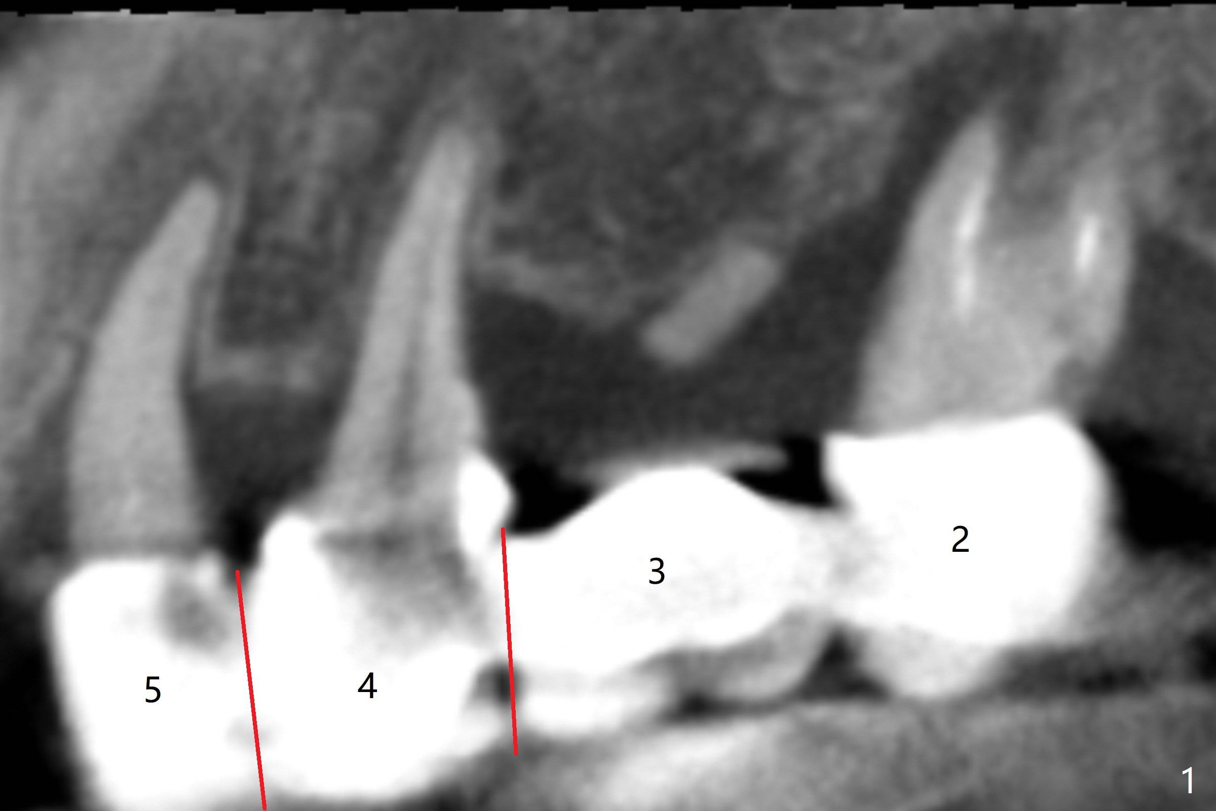
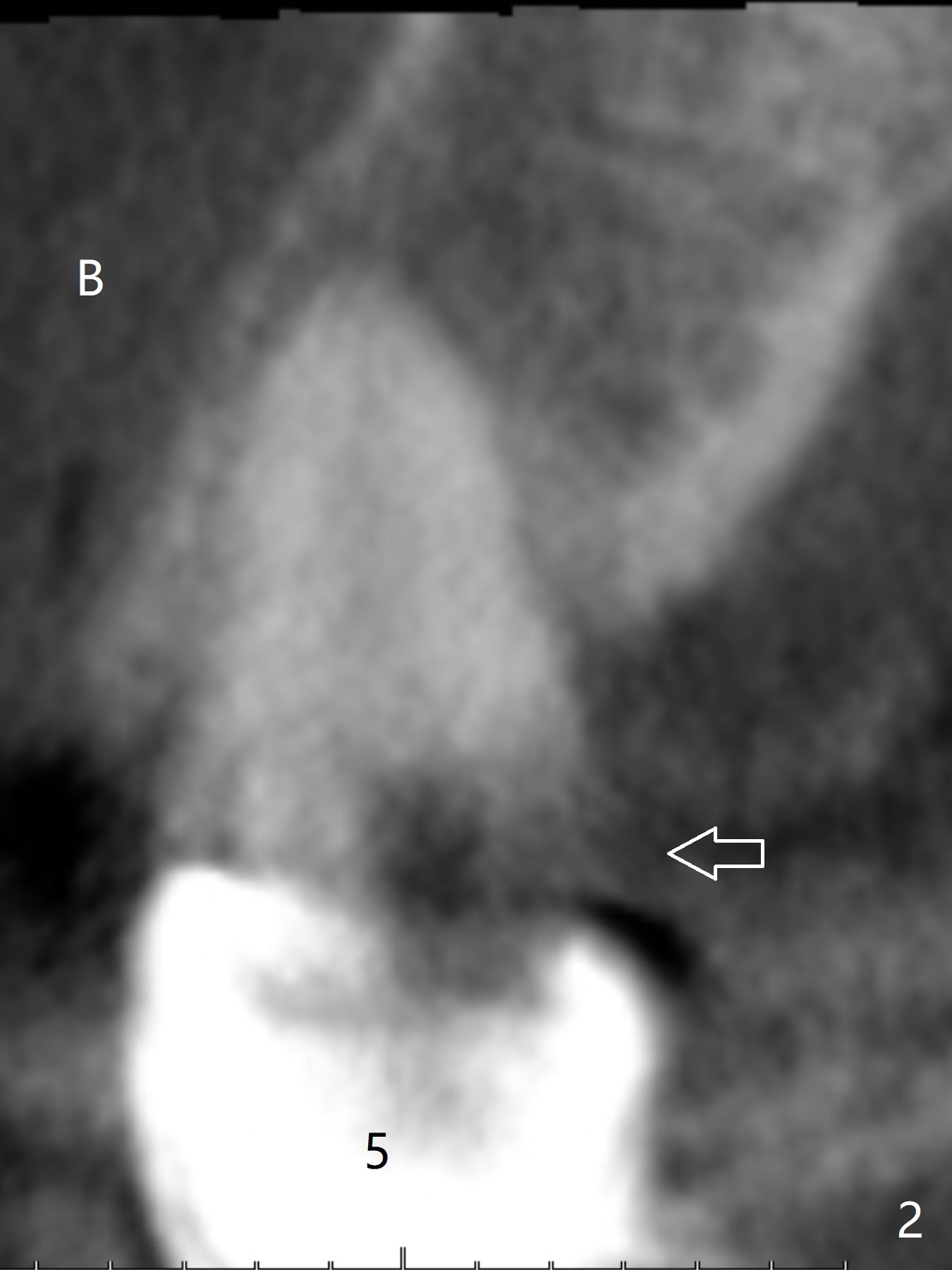
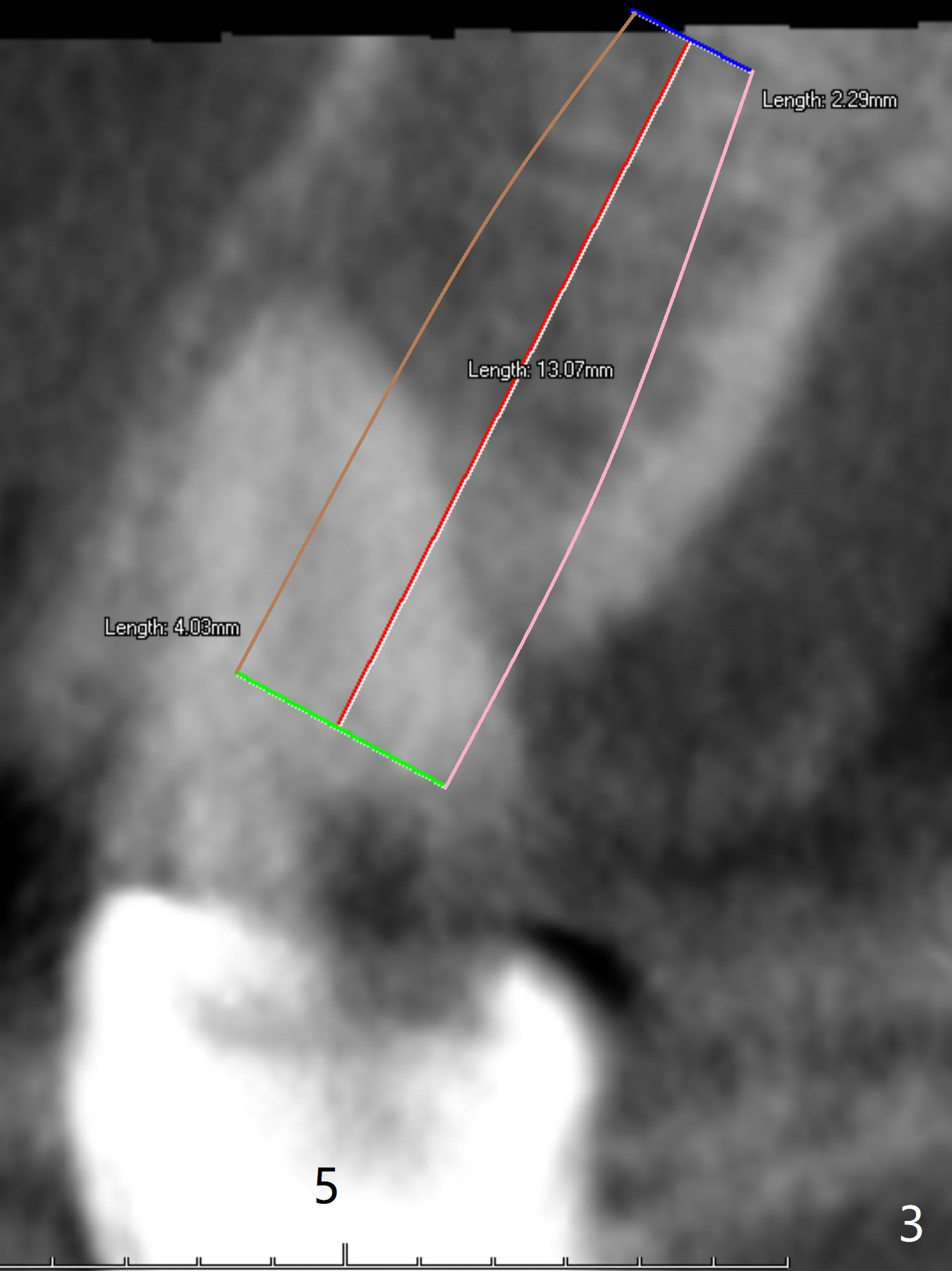
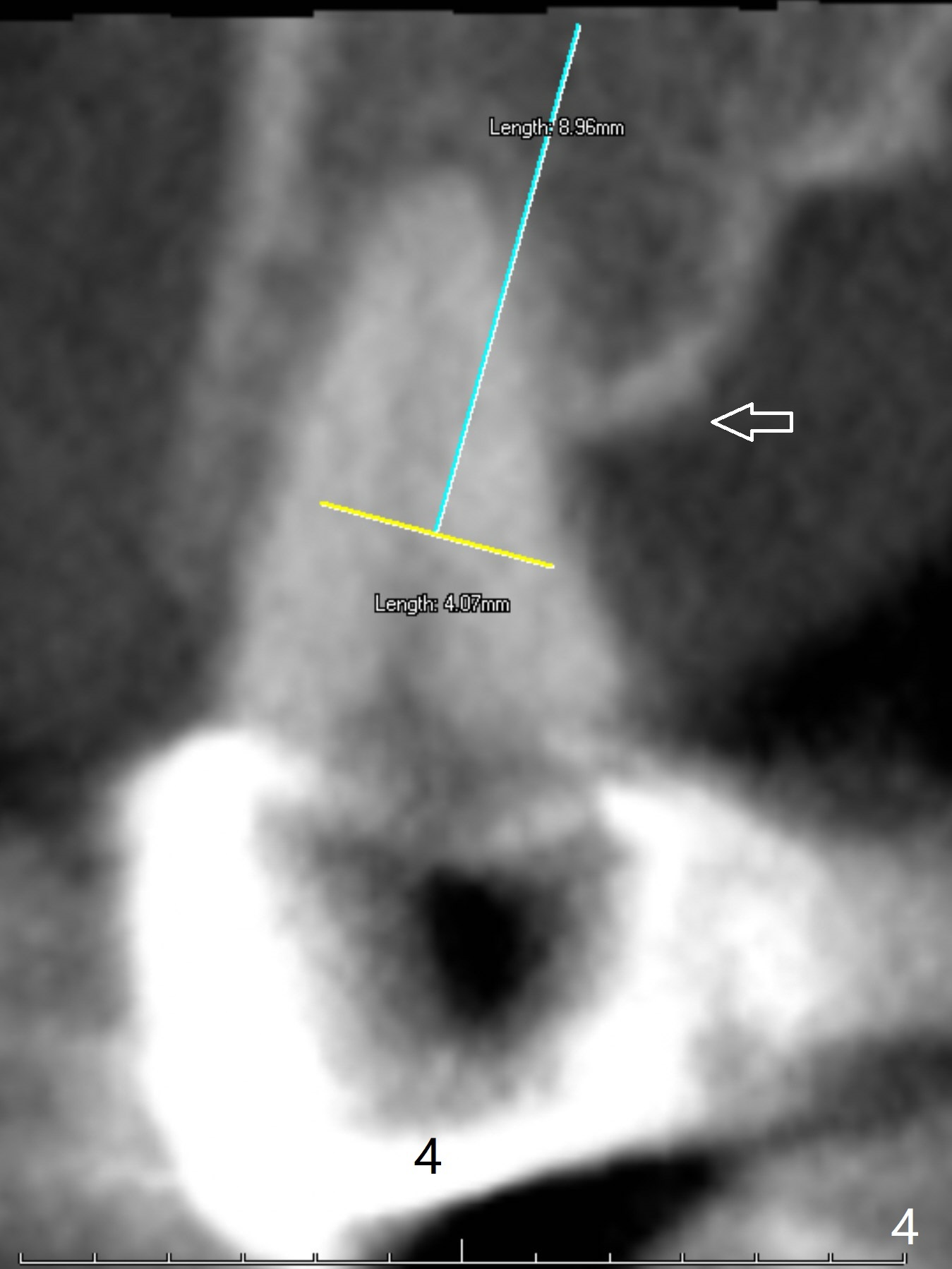
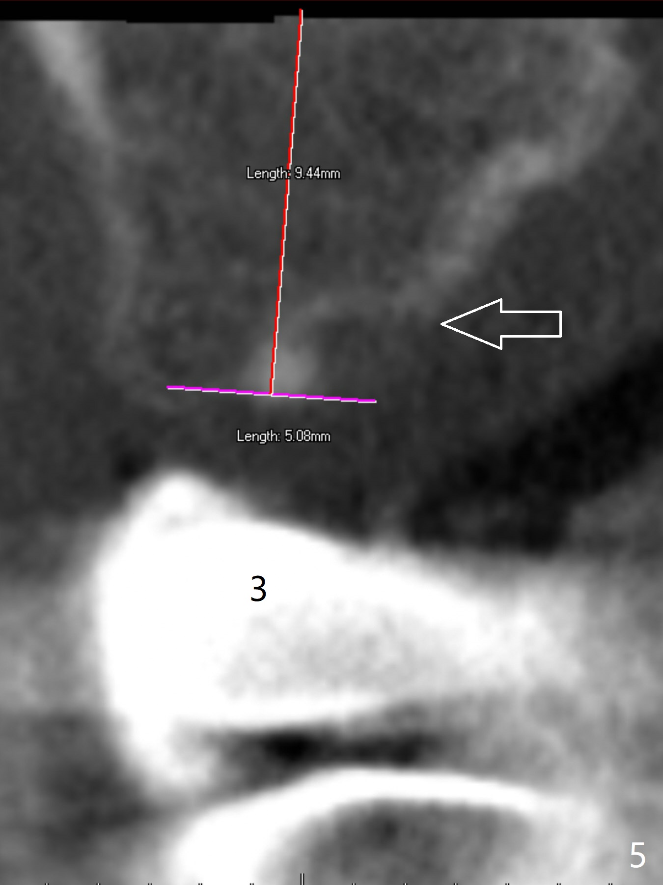
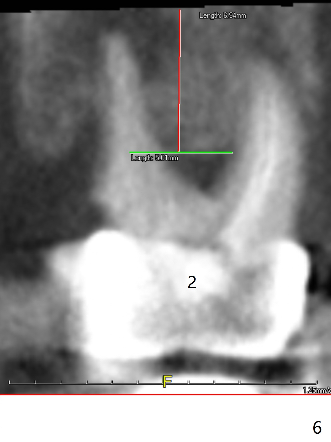
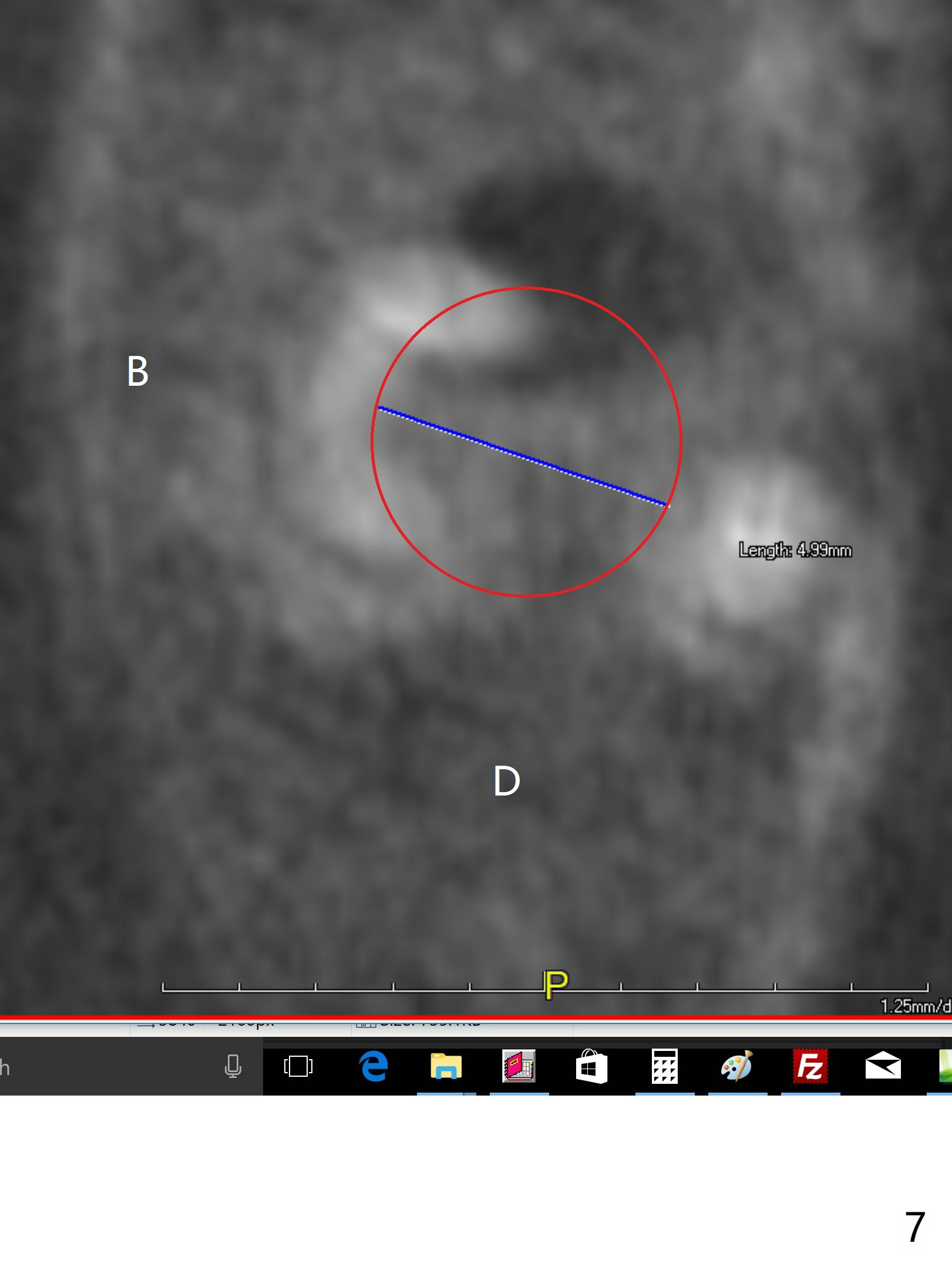
A 43-year-old woman has a failing upper right bridge (Fig.1: #2-5). While the abutment at #5 has apparently palatal open margin (Fig.2,3) and that at #4 has severe palatal bone loss (Fig.4 arrow), that at #2 has the poorest prognosis (Fig.6,7). Panoramic X-ray or PAs will be taken because of CBCT cone cut when the patient returns for #30 and 31 post-implant follow-up. Alginate impression will be taken for the upper right quadrant for provisional. The bridge will be sectioned between #3 and 5 (Fig.1 red lines) to determine salvageability of the abutments at # 4 and 5. If the latter are ok, the abutment at #2 will be extracted (expected to be loose) and implants will be placed at #2 and 3. If #4 is bad while #5 is ok, the former will be extracted and implants will be placed at #2 and 4 with a bridge. If #5 is bad while #4 is ok, implants will be placed at #2, 3 and 5.
Return to
Upper
Molar Immediate Implant, Prevent
Molar Periimplantitis (Protocols,
Table),
IBS,
Metronidazole
Xin Wei, DDS, PhD, MS 1st edition 09/23/2017, last revision 08/15/2018