,%20bone%20graft%20distal_t.jpg)
|
|
|
|
|
|
|
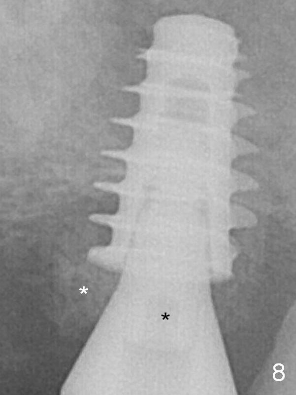
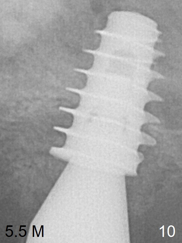
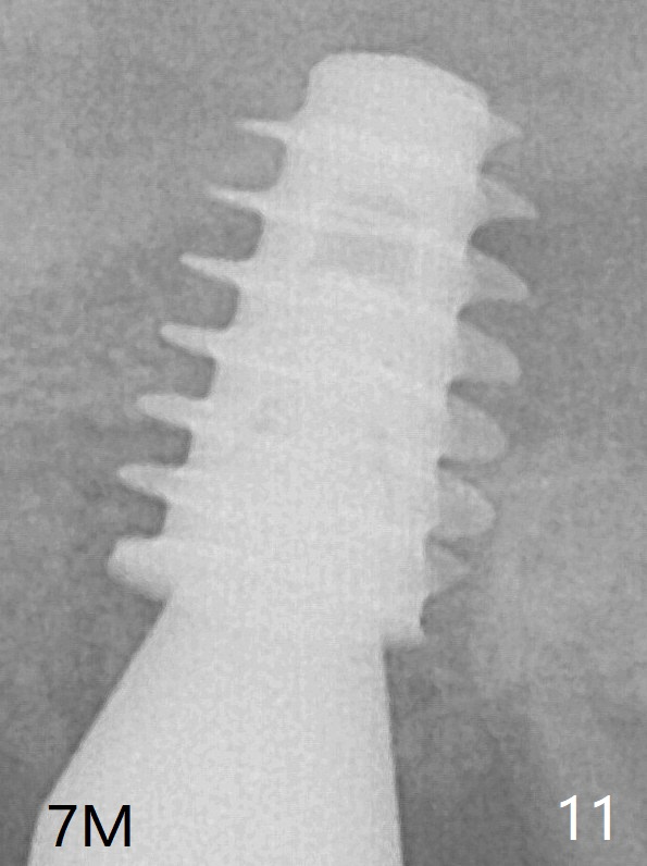
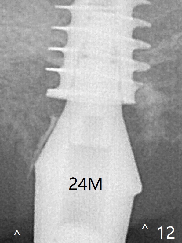
After placement of PRF membrane and bone graft (allograft, autogenous bone and Osteogen, Fig.4 *), a 6x9 mm IBS implant is placed with insertion torque of 30 Ncm. It appears that the fins of the implant slice into the bone at high magnification for engagement. Following further placement of the implant, bone graft is packed into the distal portion of the socket (<). The mesial gingiva is so thick that it is impossible to place bone graft mesial to the implant. When an implant is placed at #13 one month later, extend the incision to the mesial of #15 for bone graft. One month later, bone graft is placed mesial (Fig.8 white *) and palatal (black *) to the implant at #15 while a 3.8x13 mm implant is placed at #13 following bone expansion (using Magic Split and Magic Expander 3.0 mm). The pair abutment is changed to a healing one (6x4 mm) because of mobility 2.5 months postop. The implant seems to be stable 5.5 months postop (Fig.10), while the bone graft stays. The implant remains stable clinically 7 months postop (Fig.11). The bone graft appears to mature and covers the abutment with provisional 24 months postop, the bony changes is related to the thick gingiva (Fig.12 ^).
Fins of IBS Implant Course 2 3 4 CMC Last Next Xin Wei, DDS, PhD, MS 1st edition 03/01/2019, last revision 03/01/2019