,%20bone%20graft.jpg)
|
|
|
|
|
|
|
|
|
|
|
|
|
|
|
|
||
|
|
|
|
||
Underprep for Senior with Soft Bone
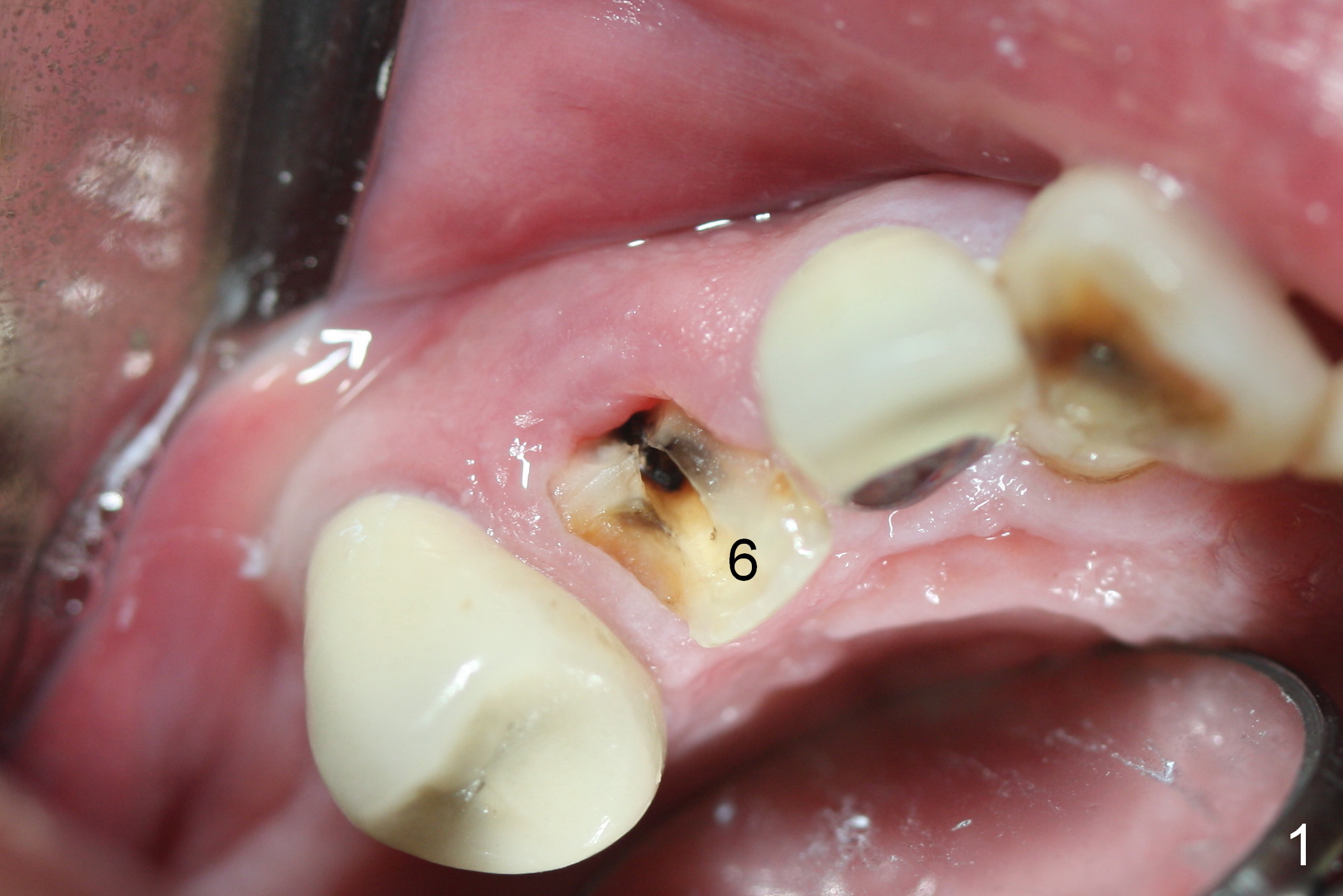
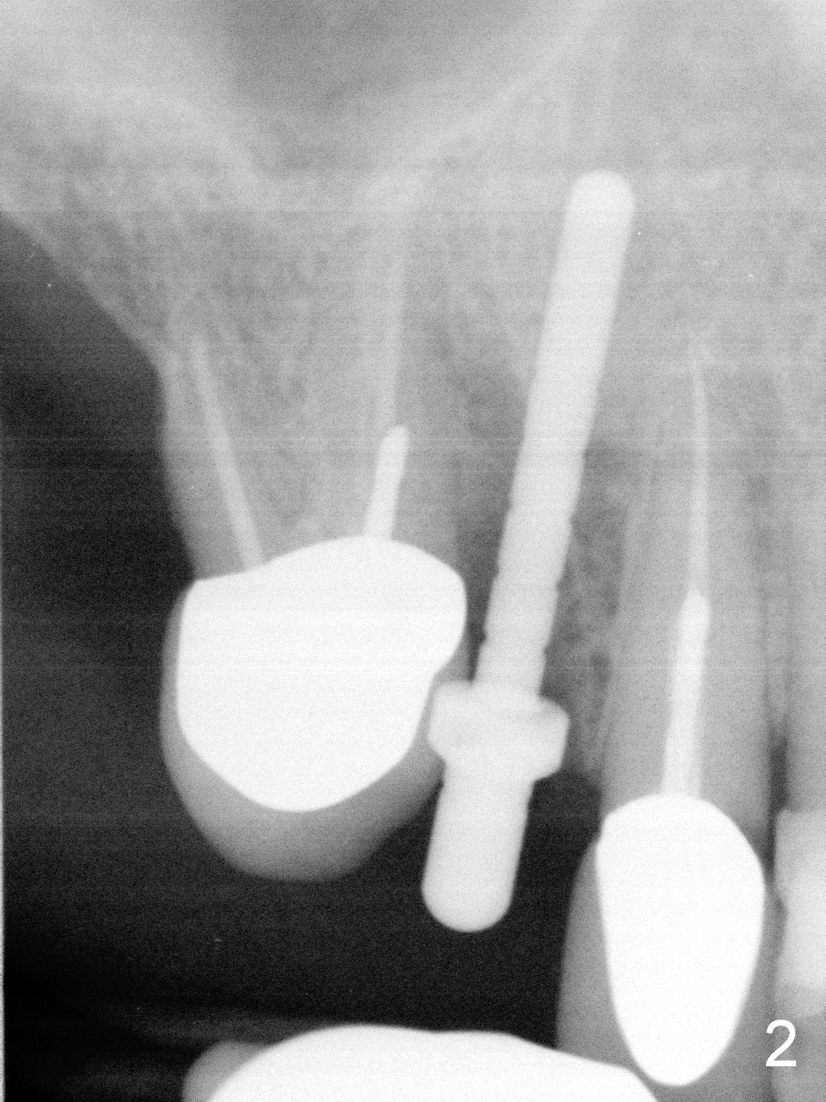
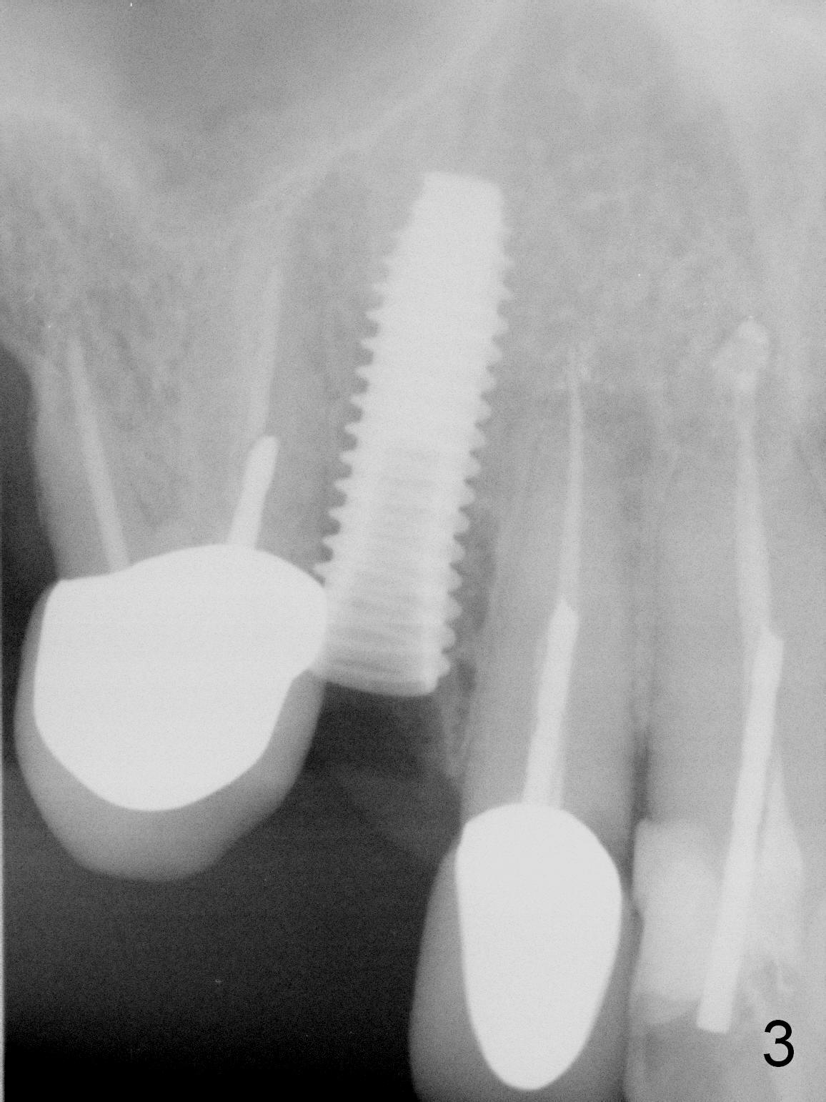
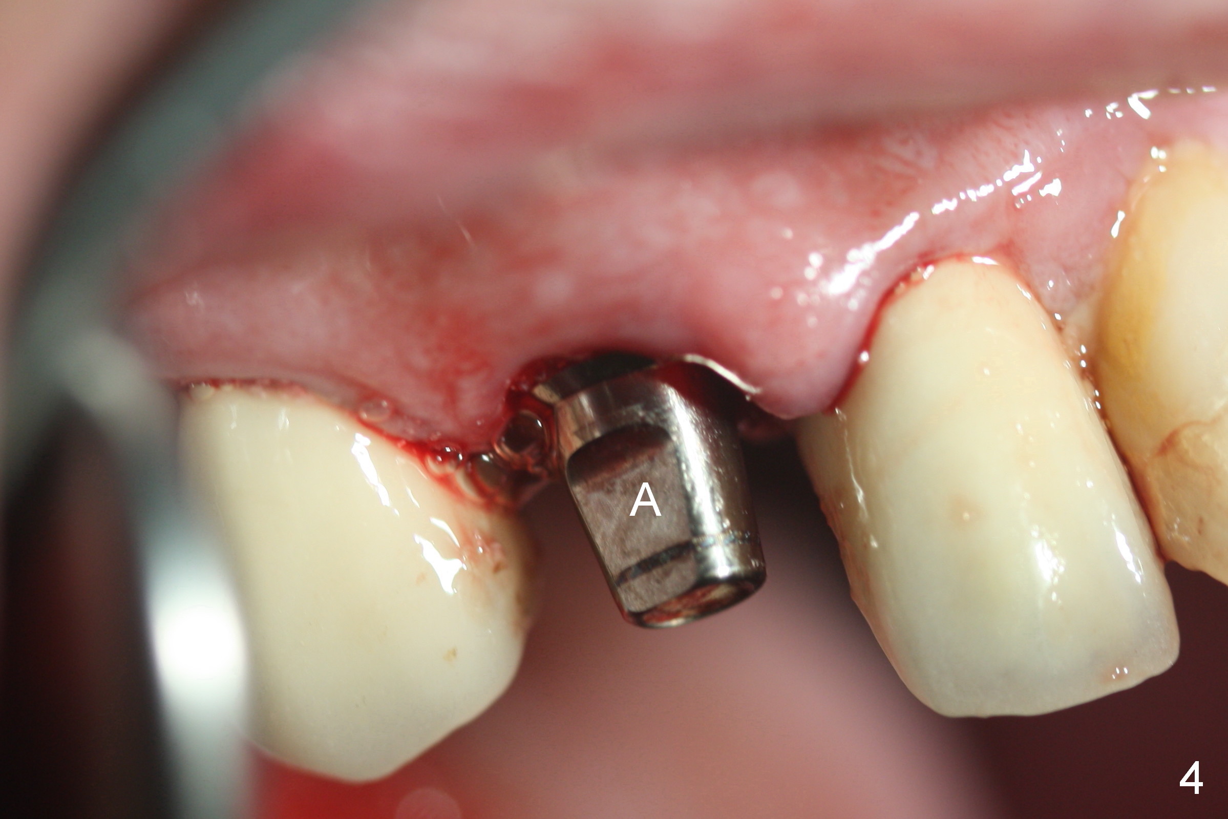
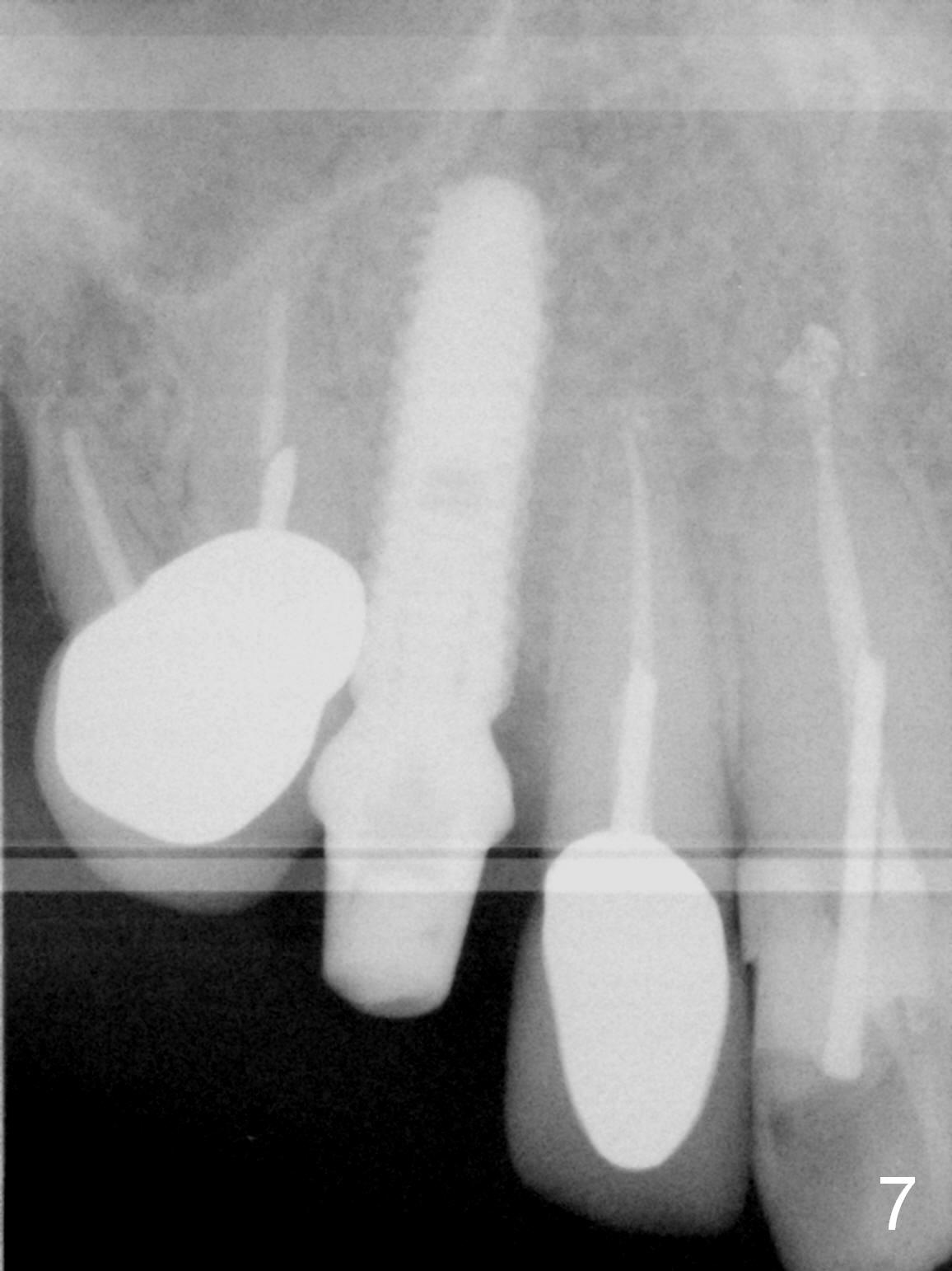
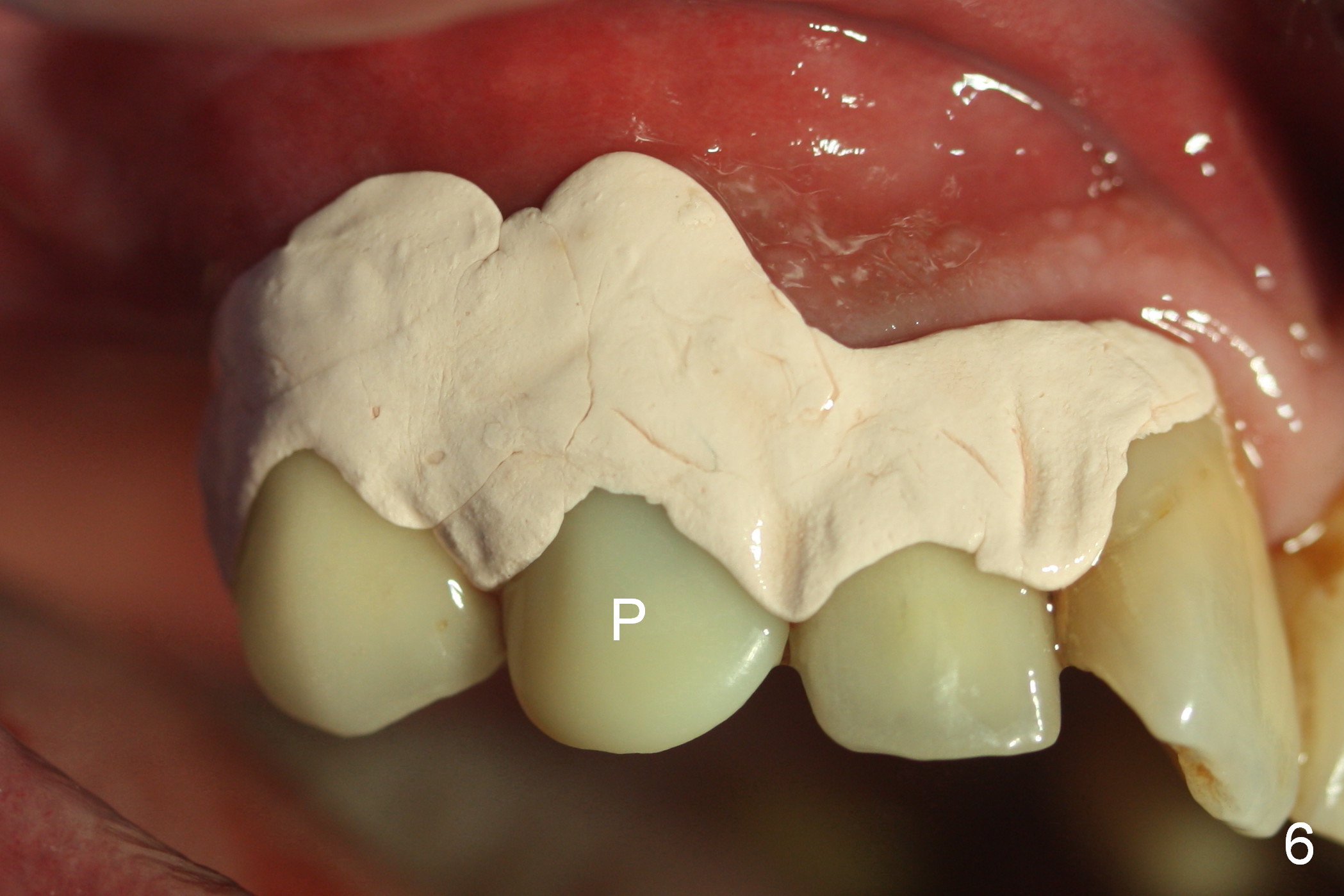
After extraction of the subgingival fractured upper right canine (Fig.1), the buccal plate is found to have been perforated at the apex. the root is measured 5x12 mm. Initial osteotomy with a 2 mm pilot drill shows that the bone is soft (Fig.2). After use of a 3.2 mm drill (underprep, normal drill size (3.7 mm)), a 4.5x15 mm implant is placed with insertion torque > 50 Ncm (Fig.3). The implant is further torqued until the implant plateau is 3 mm apical to the buccal gingival margin; a 5.5x5(3) mm abutment is placed (Fig.4,5 A). Osteogen plug is inserted into the apex of the socket, while the rest of gap is filled with mineralized cortical allograft and Osteogen. An immediate provisional (Fig.5,6 P) is fabricated to keep the graft in place, followed by periodontal dressing (Fig.6).
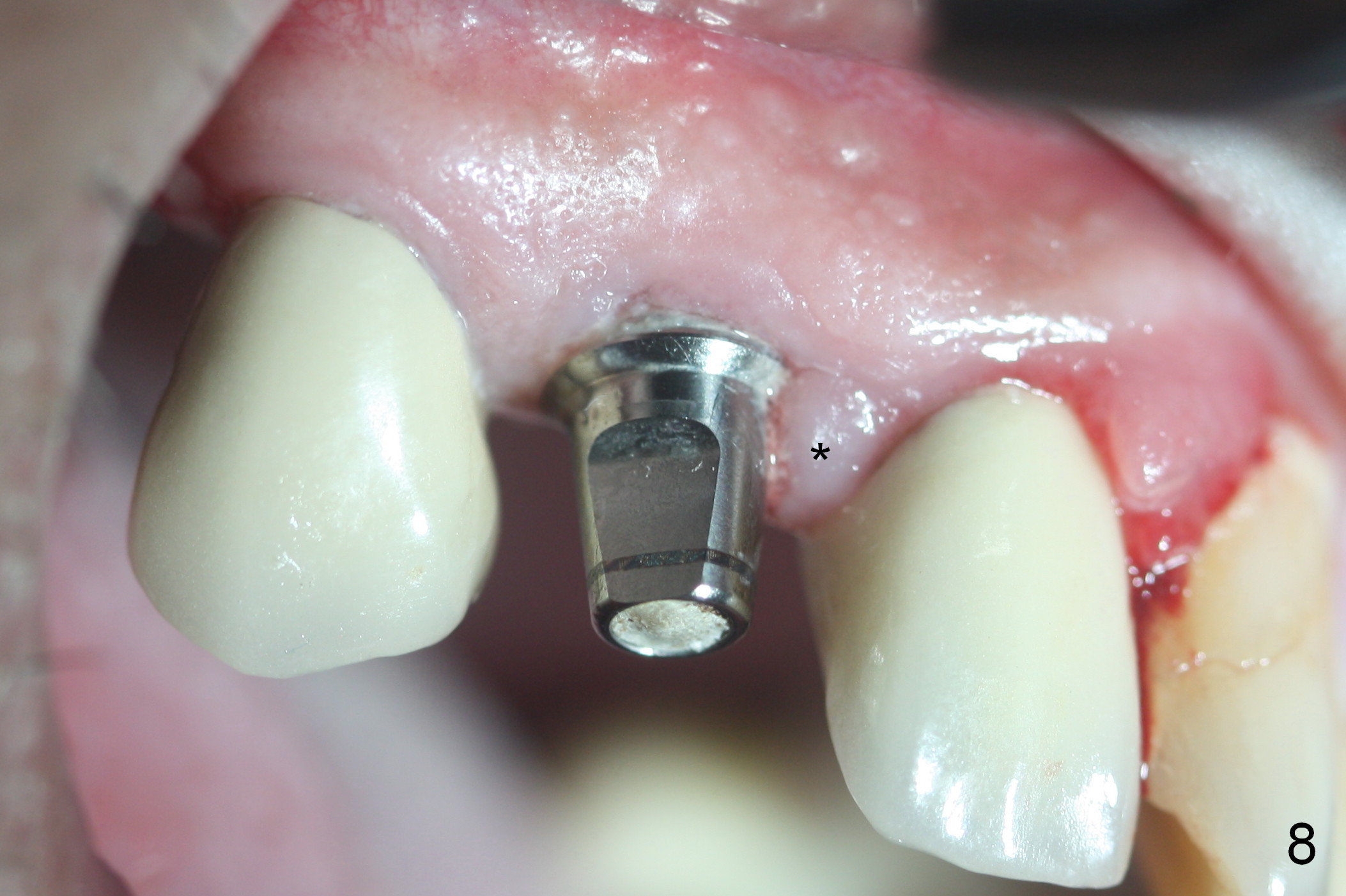
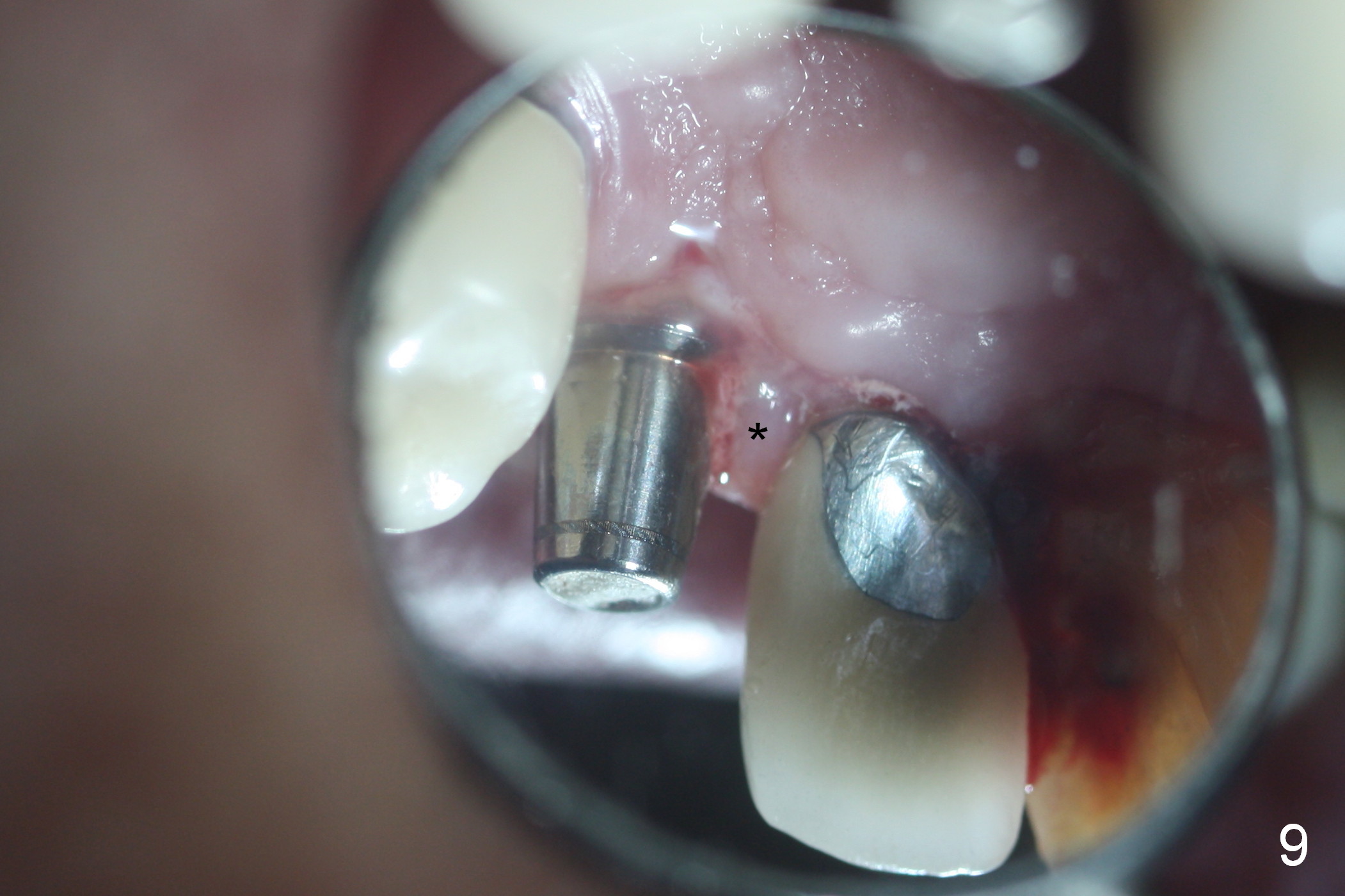
Three months postop, the provisional is dislodged (Fig.7-9). The mesial gingiva is hypertrophic buccally (Fig.8 *) and lingually (Fig.9 *). There appears to biologic width violation (Fig.5). The abutment should be changed to the one with smaller in diameter and longer in cuff (4.5x5(4) mm) with the buccal margin reduction.
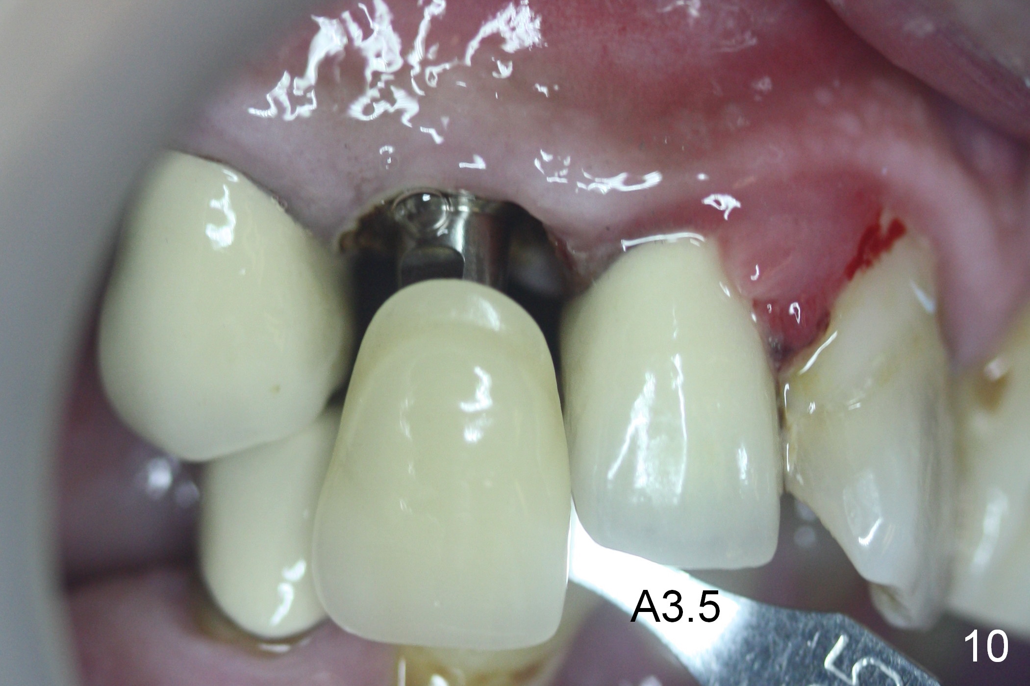
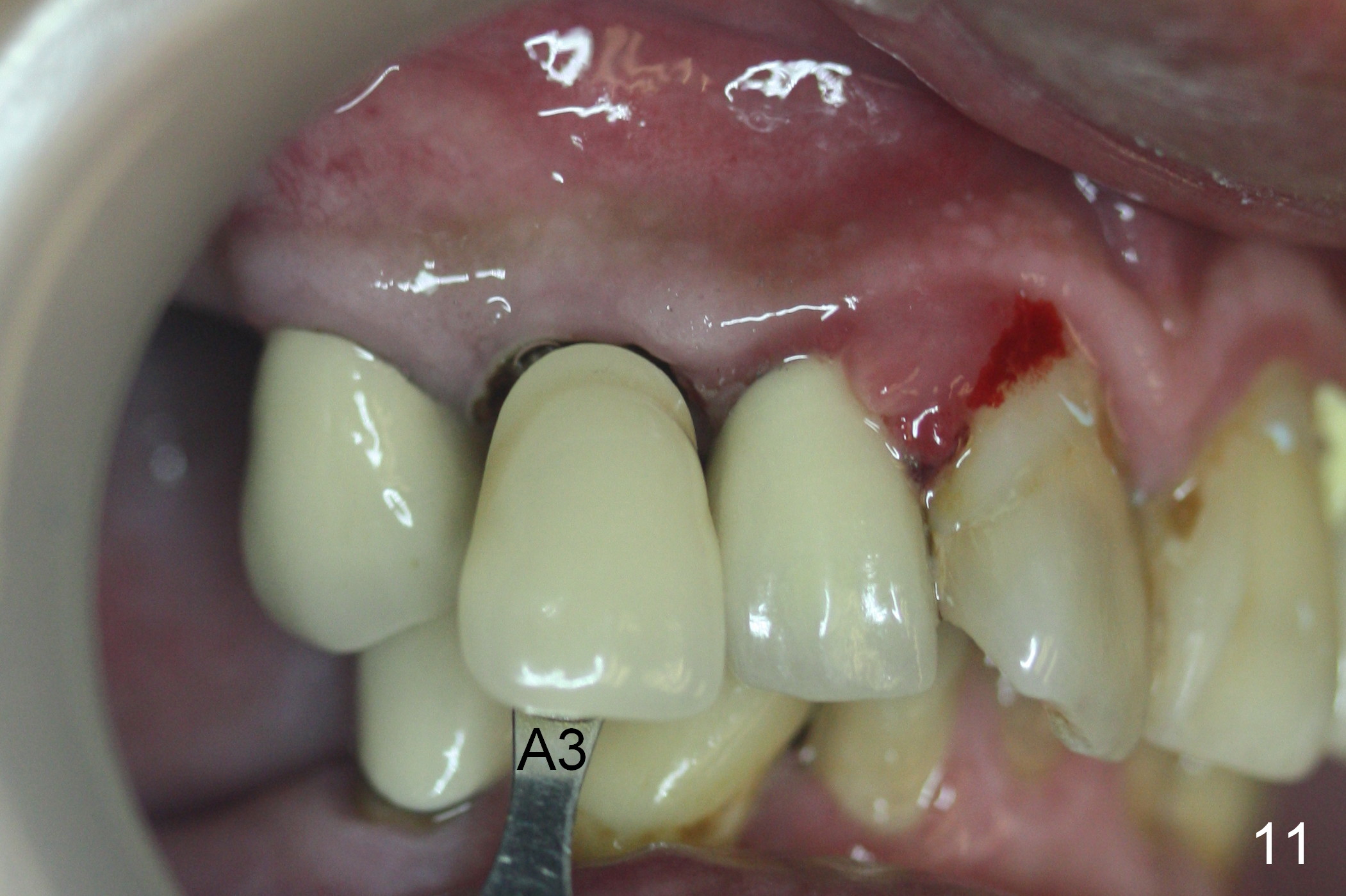
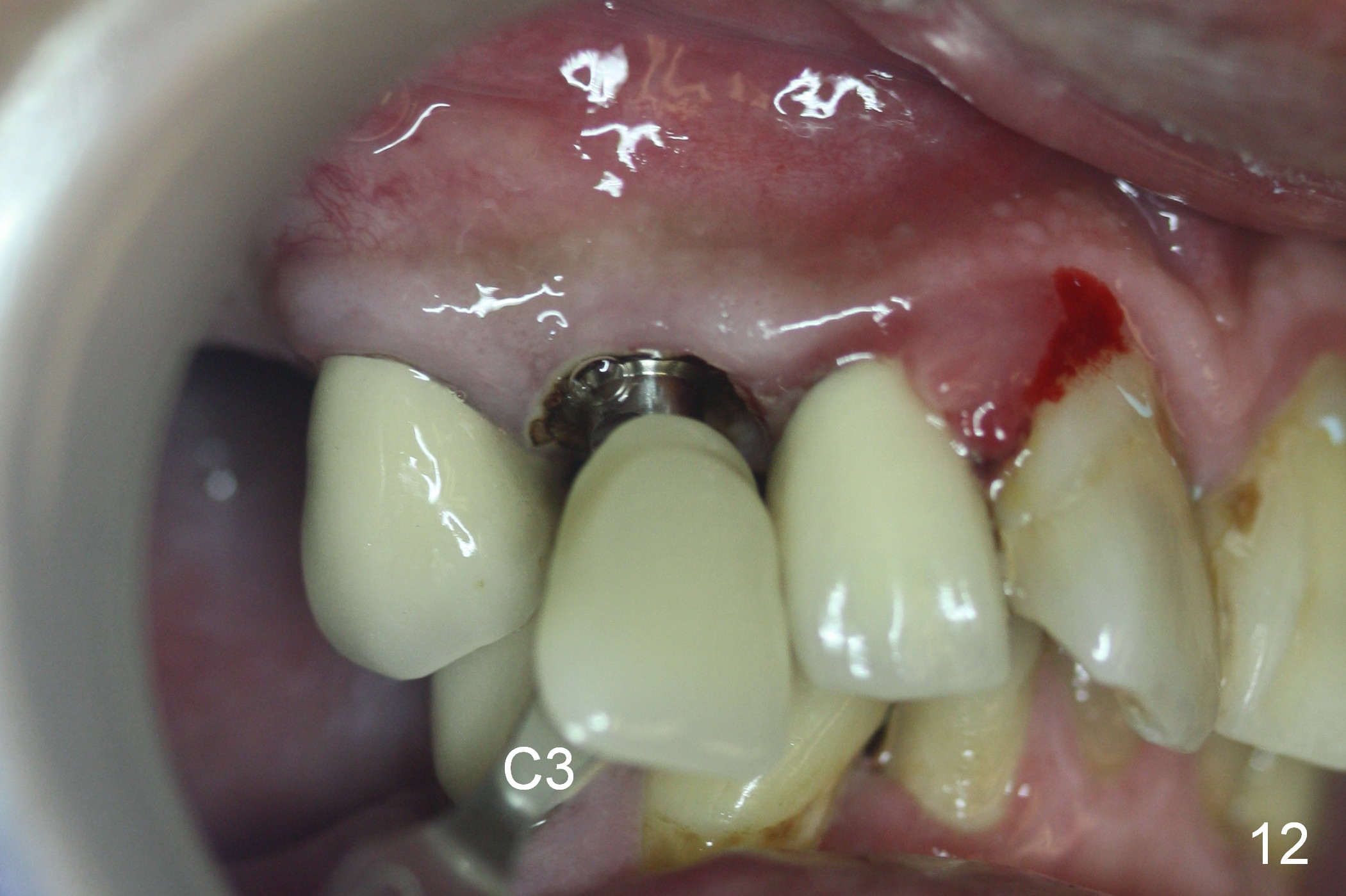
Six days post provisional reline, the mesial papilla erythema has subsided substantially; the abutment is changed to 4.5x5(2) with Diode gingivectomy (Fig.10-12).
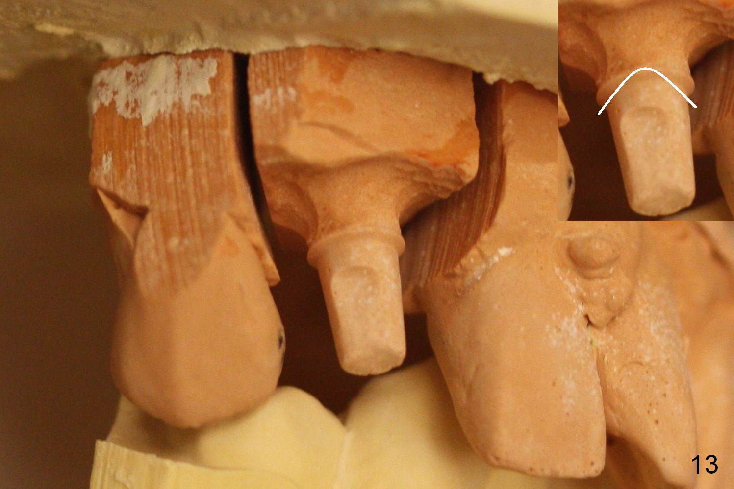
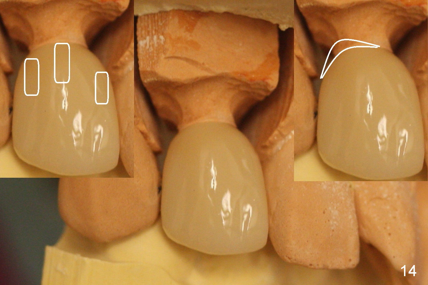
In fact the abutment margin is flat (Fig.13, not trimmed as shown in the inset). To avoid excess cement, cement is applied short of the mesial margin inside the crown (Fig.14 left inset (white area)). Once the crown is seated with cement, the excess one is present anywhere other than the mesial (Fig.14 right inset (white area)).
Return to
Upper
Canine, Upper Arch Immediate Implant,
Professionals,
Technicians
Xin Wei, DDS, PhD, MS 1st edition 01/15/2016, last revision 08/30/2016