

 |
 |
 |
 |
|
 |
 |
 |
 |
|
 |
 |
 |
||
 |
 |
 |
||
Incisor or Canine?
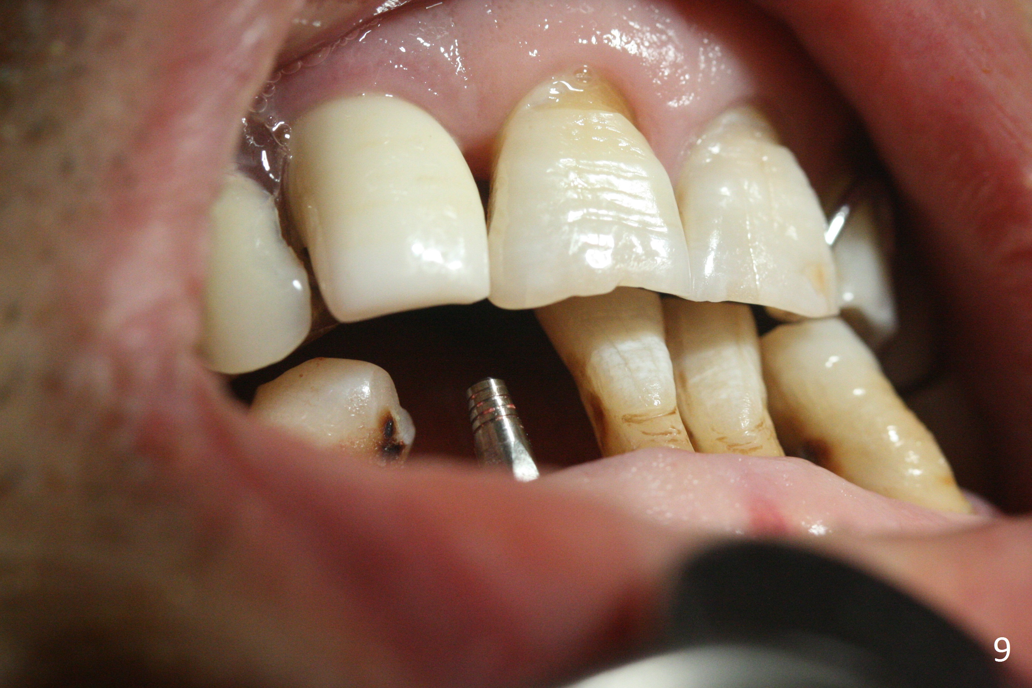
The lower dentition is special, consisting of a residual root (Fig.1 ^), 2 incisor (I), 1 canine (C), 1 premolar (P) and 1 molar (M). The residual root looks like an incisor with rotation of 90º (Fig.2,3). Osteotomy is initiated (Fig.4) for a 3x16(2) mm 1-piece implant (Fig.5 with 45 Ncm). The implant is being placed as distal as possible (Fig.4 arrow) so that a large canine-like provisional is to be fabricated in the large edentulous space (Fig.8,9) after bone graft (Fig.6,7 *). The gingiva around the provisional (Fig.10 P) remains healthy 11 days postop with occlusal clearance against the opposing dentition (Fig.11). The implant threads are not exposed with the help of bone graft 3 months 1 week postop (Fig.12). The gingiva around the implant is healthy (Fig.13). Soft tissue socket is formed by the provisional (Fig.14 *).
Return to
Lower
Incisor,
Canine Immediate Implant,
IBS,
Shield
Xin Wei, DDS, PhD, MS 1st edition 03/31/2017, last revision 02/20/2021