|
|
|
|
|
|
|
|
Thickness and Height of the Buccal and Lingual Plates of the Lower Bicuspid
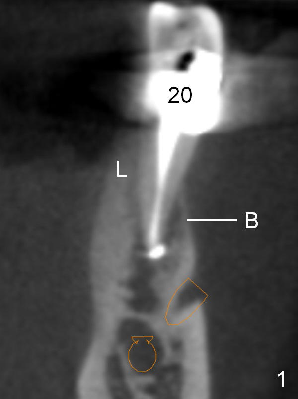
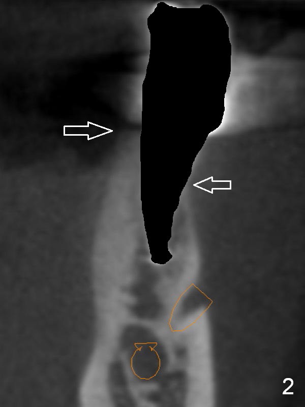
Fig.1 is a CBCT coronal section of the lower left bicuspid of a 57-year-old lady. The buccal (B) bone is thin and mainly cancellous, while the lingual (L) bone thick and almost exclusively cortical. When the tooth is schematically extracted, the lingual plate is higher than the buccal one (Fig.2 arrows).
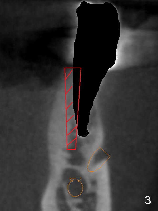
If osteotomy follows the path of the socket, an implant will be most likely deviated buccally, creating restorative difficulty.
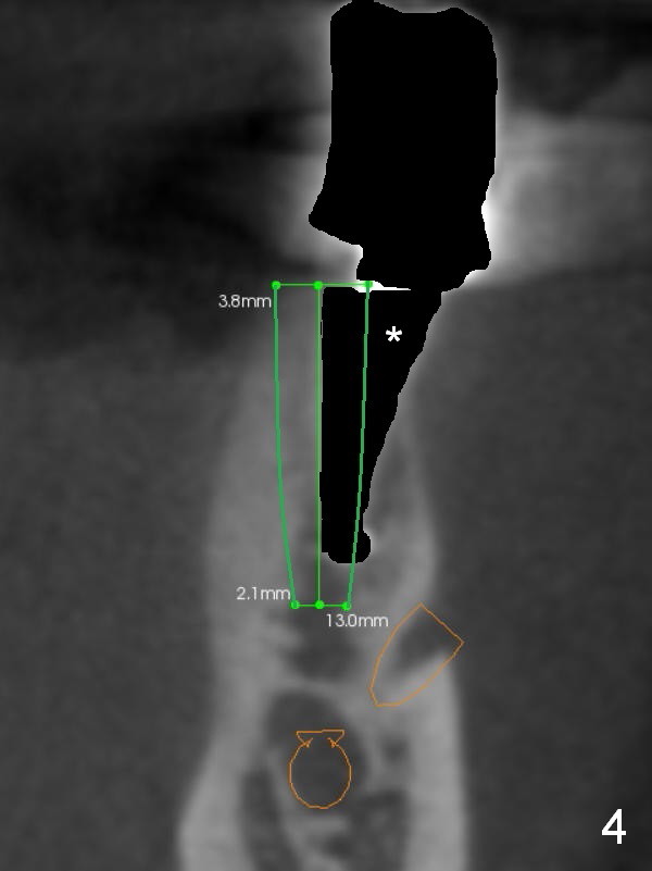
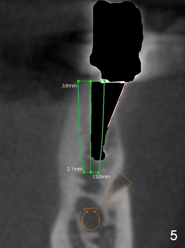
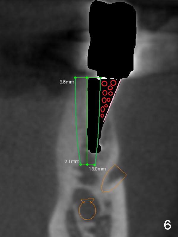
When the osteotomy is intentionally placed as lingual as possible (Fig.3 red area), the implant (Fig.4 green) will end up in a more favorable position with a large buccal gap (*). The latter allows to place collagen membrane (Fig.5 pink) and bone graft (Fig.6 red circles).
Return to Lower Premolar Immediate Implant
#28
Xin Wei, DDS, PhD, MS 1st edition 12/27/2015, last revision 01/19/2018