|
|
|
|
|
|
|
|
|
|
||
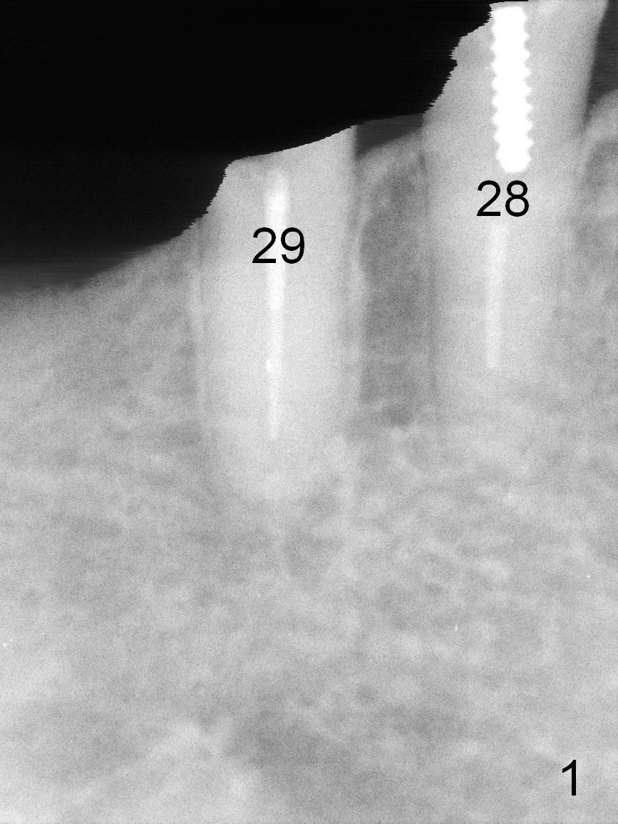
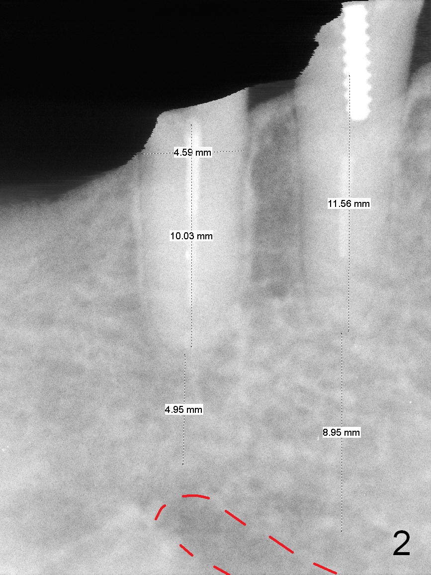
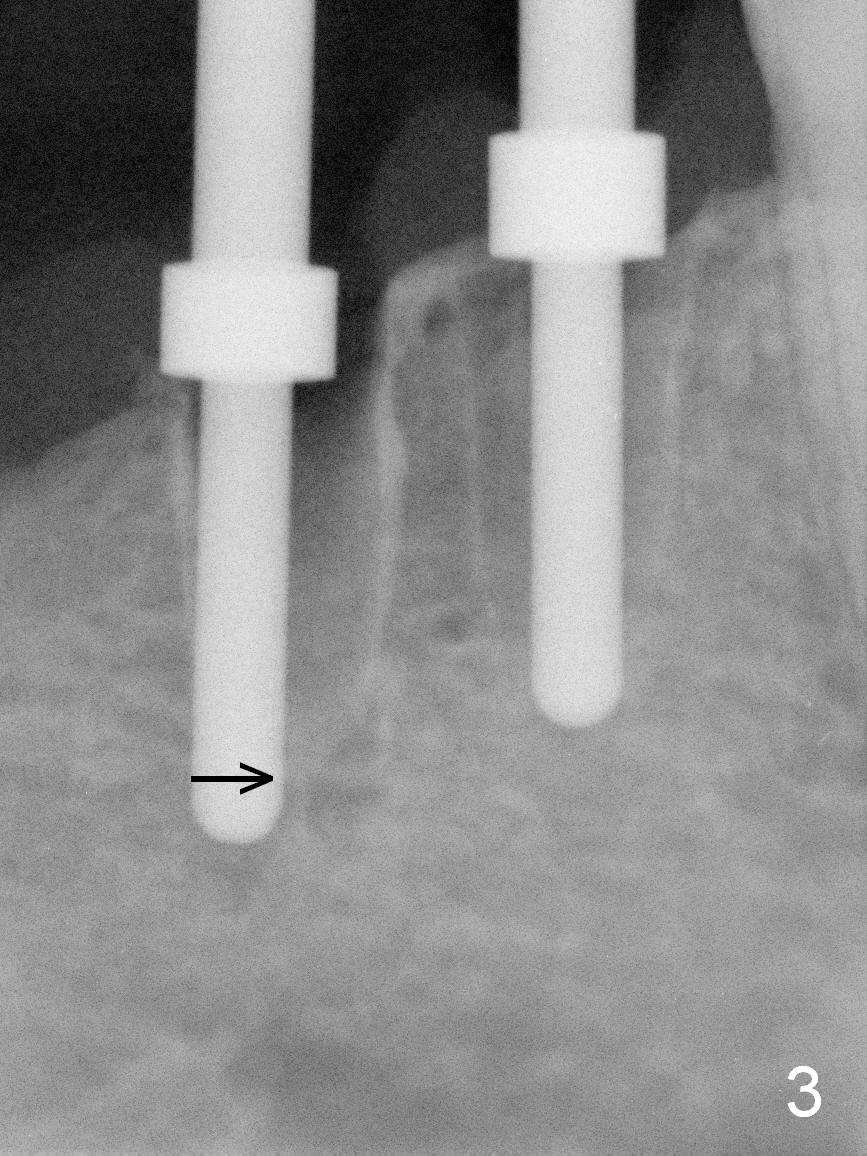
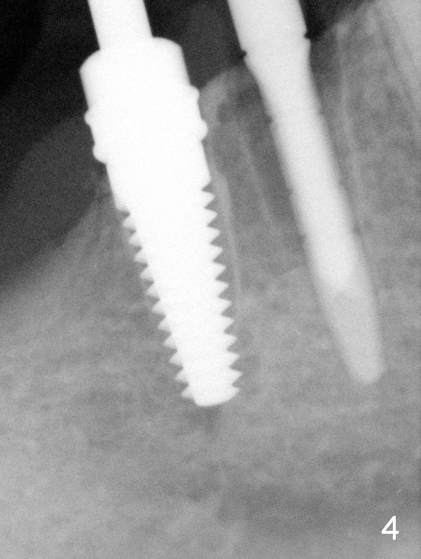
Osteotomy Depth of Lower Premolars
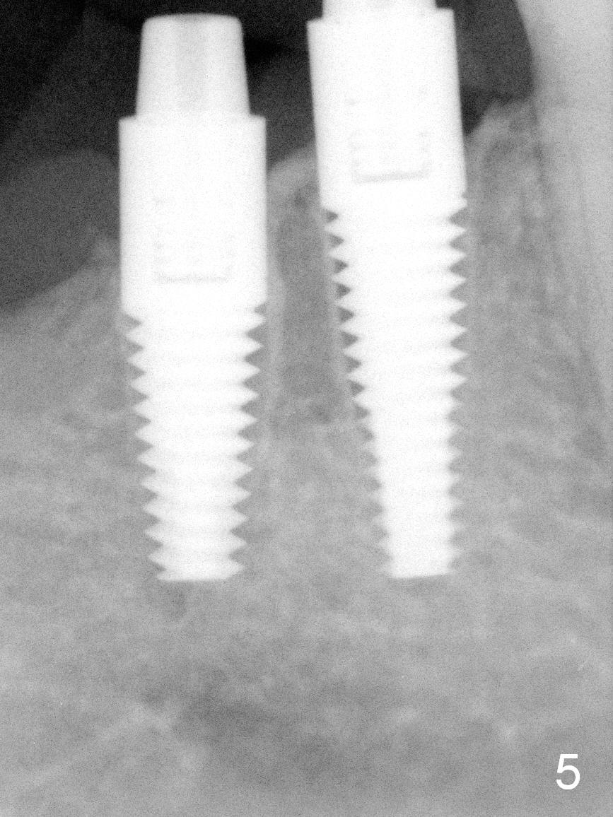
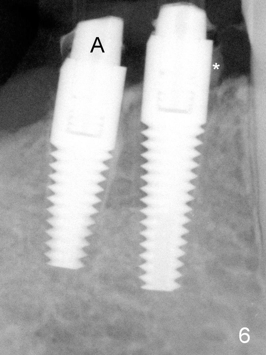
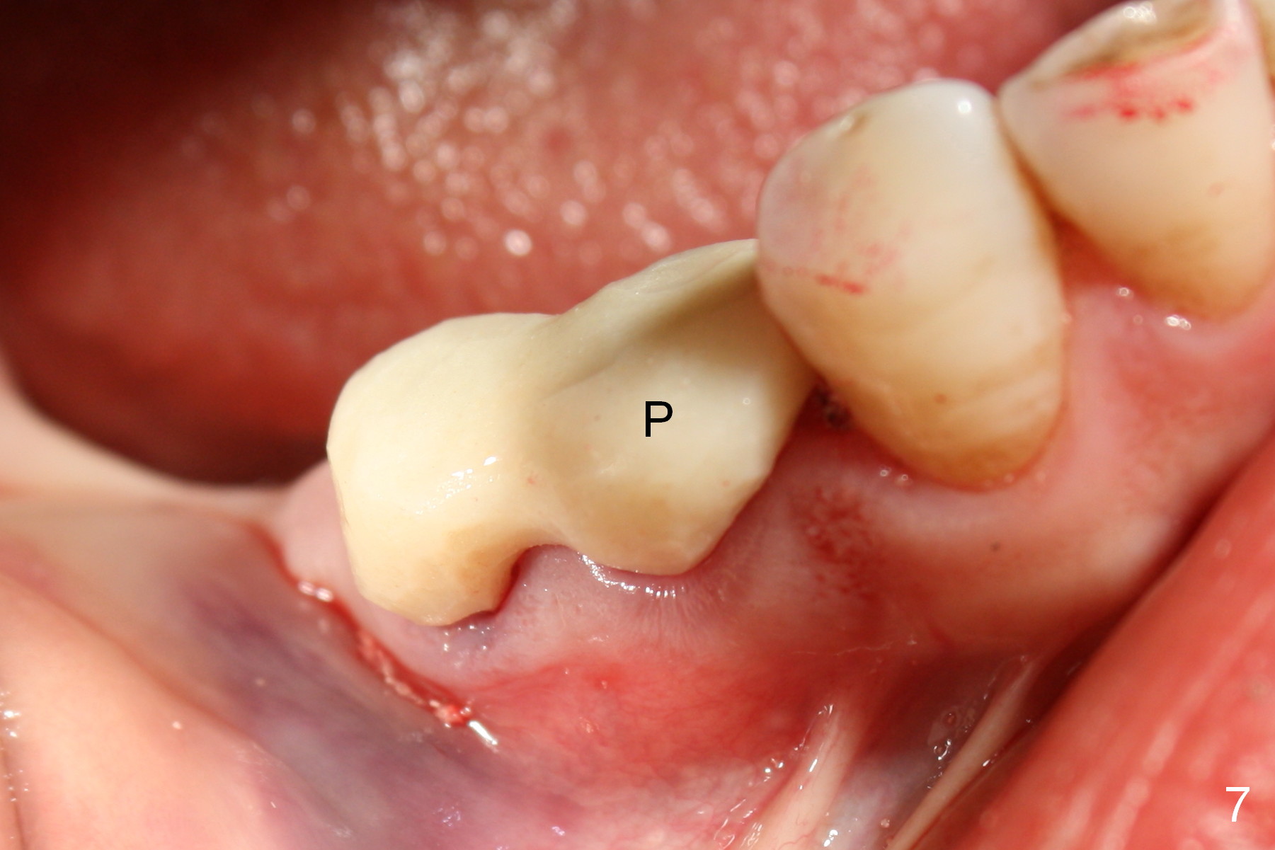
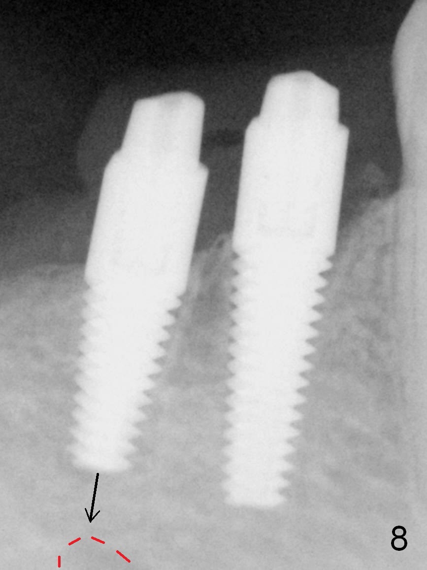
After restoring the implant at the site of #20, the patient returns for #28,29 implant placement (Fig.1,2). Bone level and distance from the Mental Loop (Fig.2 red dashed line) are different. It appears that longer implant can be placed at #28 than that at #29. Parallel pins are placed after initial osteotomy (Fig.3), it appears that the osteotomy at #29 should be moved mesially (arrow). Next PA shows that the position of the osteotomy at #29 is corrected (Fig.4). The position of the implants (4.5x17, 4.5x14 mm) appears ideal (Fig.5). After preparation for an immediate provisional, bone graft is placed in the remaining socket space (Fig.6 *). The splinted provisional is temporarily placed (Fig.7 P). The implant at #29 seems to be buccally placed. After CBCT confirmation, it should be removed for replacement. In addition to moving the osteotomy lingually, there is apparent space (~3 mm) to extend the osteotomy apcially for primary stability (Fig.8 arrow). The apical diameter of the implant is 3 mm.
Return to Lower Premolar Immediate Implant Xin Wei, DDS, PhD, MS 1st edition 09/27/2015, last revision 04/04/2018