,%20trimmed%20DL,%20graft,%20Berkerly.jpg)
|
|
|
Bone graft is packed around the implant in layers after placement of a 7.5x4(5) mm abutment (Fig.3). The 1st one is composed of autogenous bone (from osteotomy) and cortical and cancellous allograft (.5-1 mm in size), while the 2nd one consists of the allograft mixed with Osteogen (alloplast). The 3rd layer is collagen plug cut in strip (gingival zone (connective tissue reformation), while the 1st 2 layers are in bone zone for bone regeneration). The latter (collagen plug) is held in place with applying provisional acrylic over the abutment and the socket opening while it is setting. Some of acrylic is pushed into undercut areas for retention. No attempt is exerted to remove the provisional. The latter will be adjusted approximately 1 month postop. It does not matter if the bone graft is placed in the gingival zone, since the bone graft may be incorporated and/or embedded into a part of the gingiva.
Why does the 1st layer of graft look so dense? The first, the native bone is hard. The second, the allograft is mineralized cancellous and cortical. The size is larger than those used before (.125-.85 or .25-1 mm).
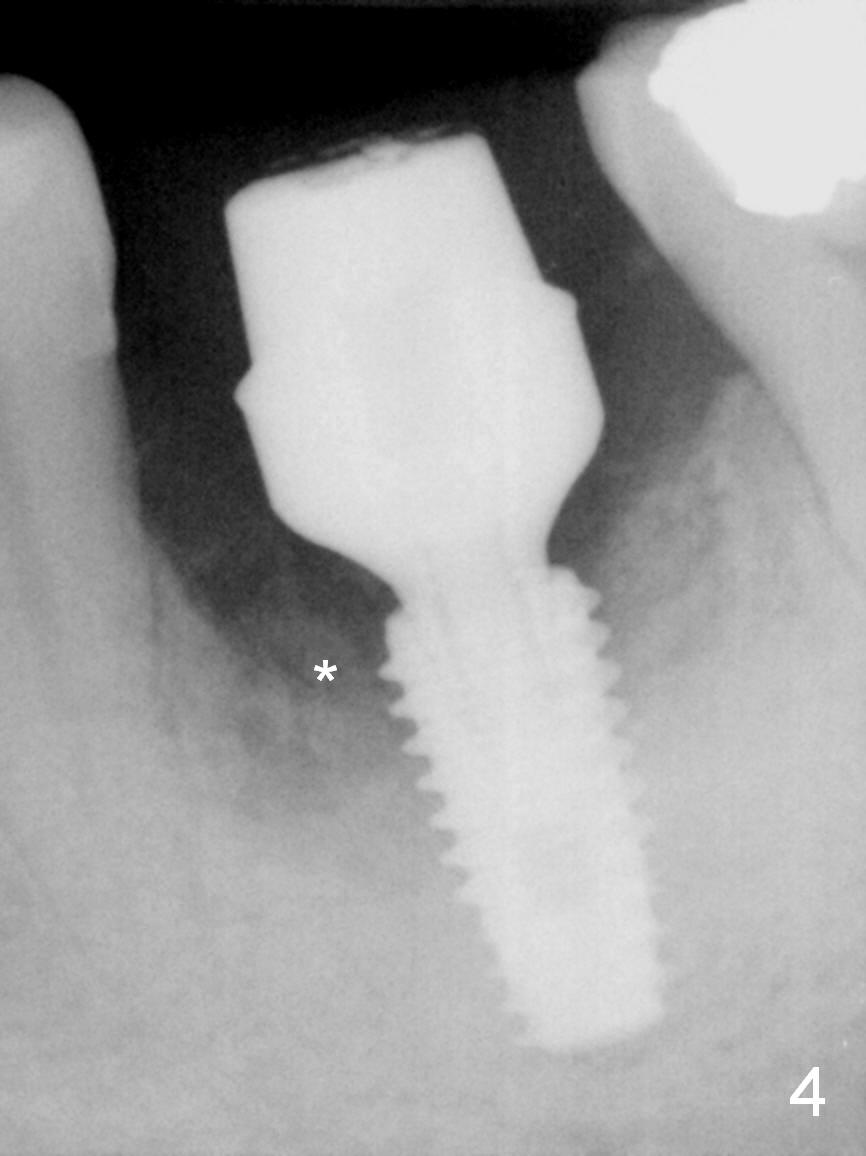
Four months postop, there is bone next to the implant (Fig.4 *). The margin of the abutment is prepared prior to impression for final restoration.
Bone Regrowth after Implant Last Next
Xin Wei, DDS, PhD, MS 1st edition 04/01/2017, last revision 01/10/2020