|
|
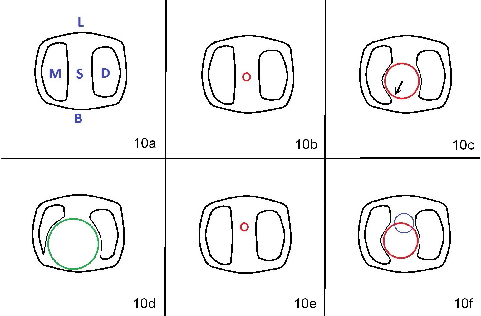
Fig.10a is an illustration of occlusal view of a lower 1st molar sockets. B: buccal; L: lingual; M: mesial; D: distal sockets; S: septum.
When the buccal and lingual plates are equal in thickness, osteotomy is initiated in the middle of the septum (Fig.10b: red circle).
When the osteotomy increases, it may deviate the least resistant area, e.g., mesiobuccally (Fig.10c arrow). Buccolingual deviation is more critical mesiodistal one, particularly apically (Fig.10d).
If the buccal plate is found to be weak beforehand, osteotomy should start more lingually (Fig.10e: red cricle).
When the osteotomy starts being deviated, correct as early as possible with a Lindamann bur and preferably over-correct (Fig.10f blue circle). The final implant placement may not encroach the buccal plate (Fig.10f red circle).
Xin Wei, DDS, PhD, MS 1st edition 02/15/2015, last revision 08/08/2015