|
|
|
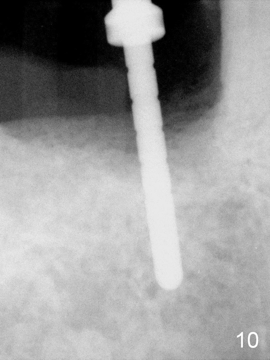
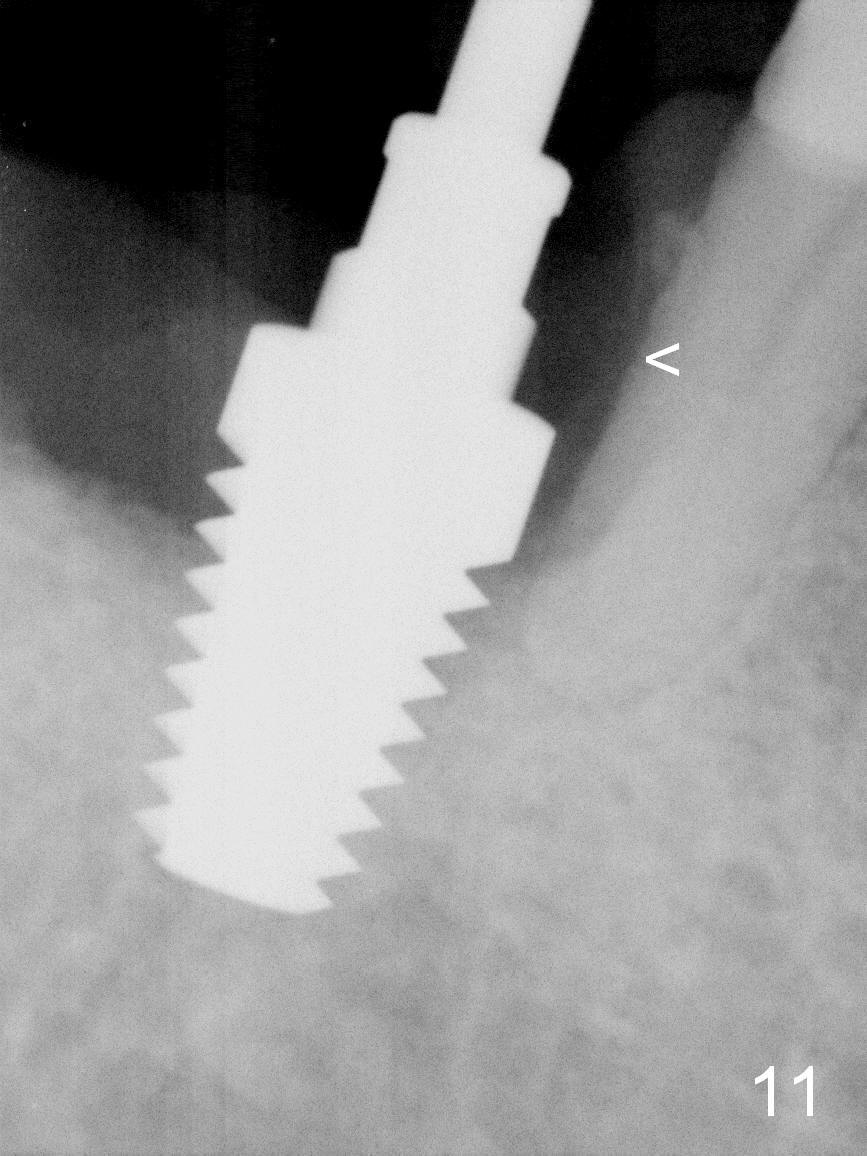
The osteotomy starts in the distolingual aspect of the mesial socket (Fig.10 (parallel pin after 2 mm pilot drill). As the osteotomy increases (using drills at 50 RPM) and is expanded (taps), the soft and hard tissue of the septum (Fig.9) is being pushed distally. Since the bone density is low and overprepartion, a 7x14 mm tap is used to obtain stability (Fig.11). The root surface of the neighboring tooth (#29) is exposed, visible from the #30 mesial socket (<). Bone graft may help repair of the bony defect.
Xin Wei, DDS, PhD, MS 1st edition 08/20/2015, last revision 08/28/2015