
|
|
|
|
|||
|
|
|
|
|
|
|
|
|
|
|
|
|
|
|
|
|
|
|
|
|
Drawback of UnderPrep
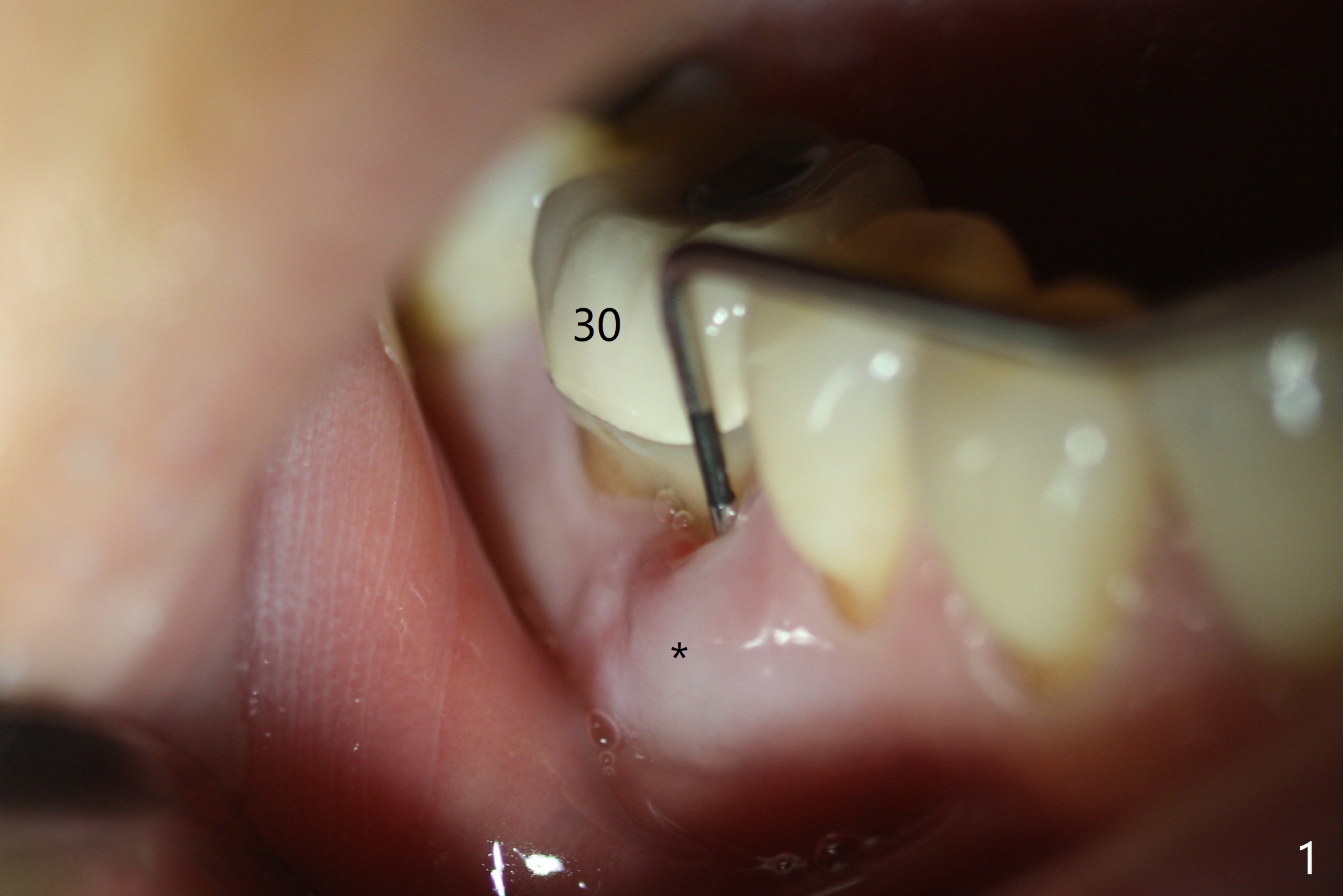
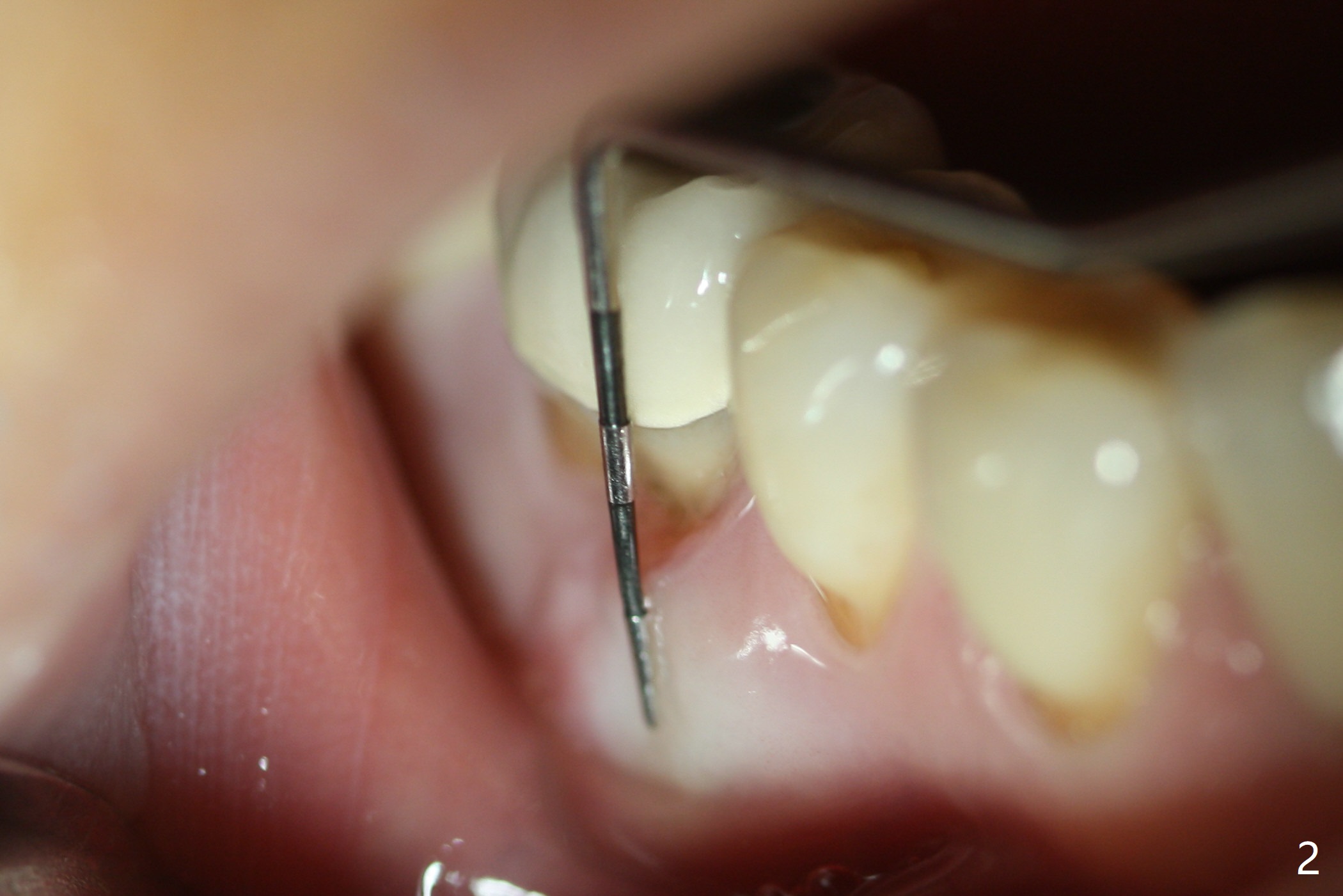
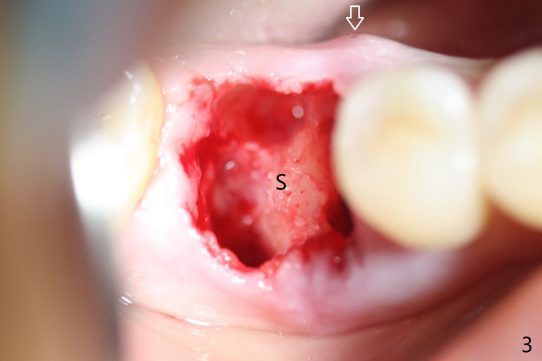
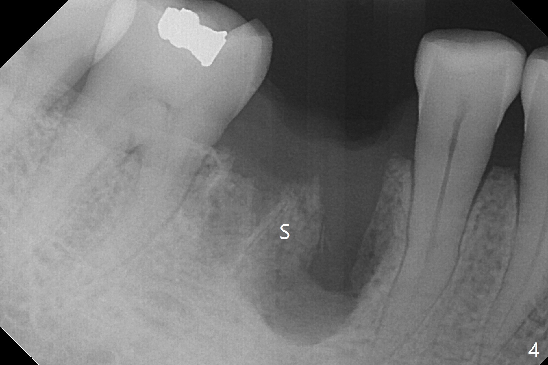
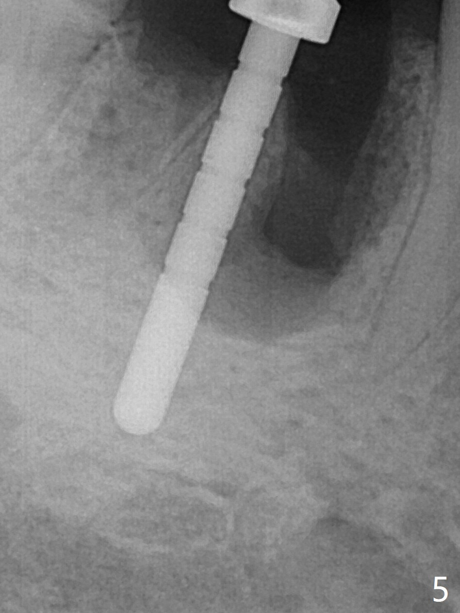
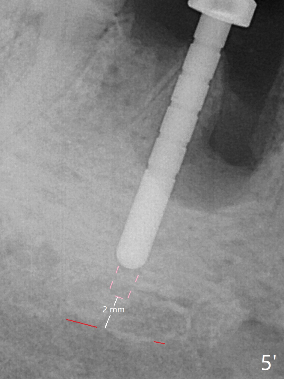
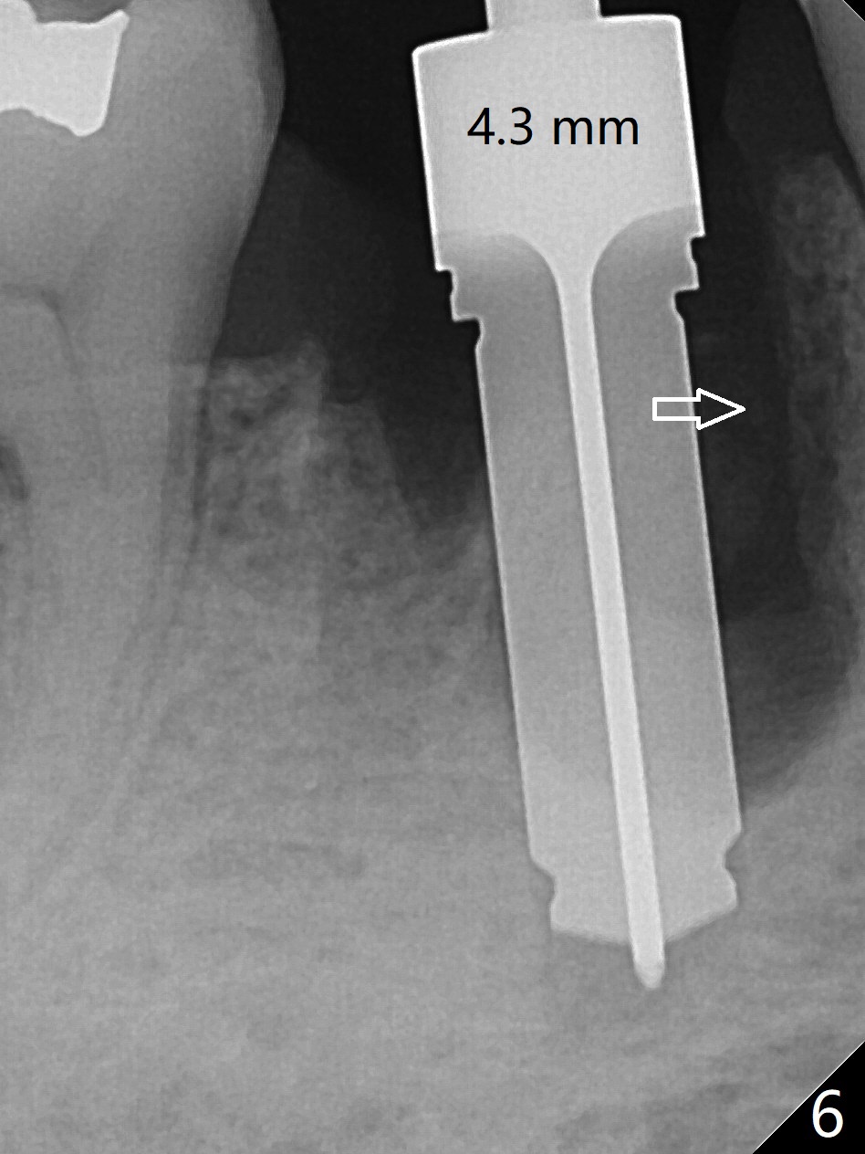
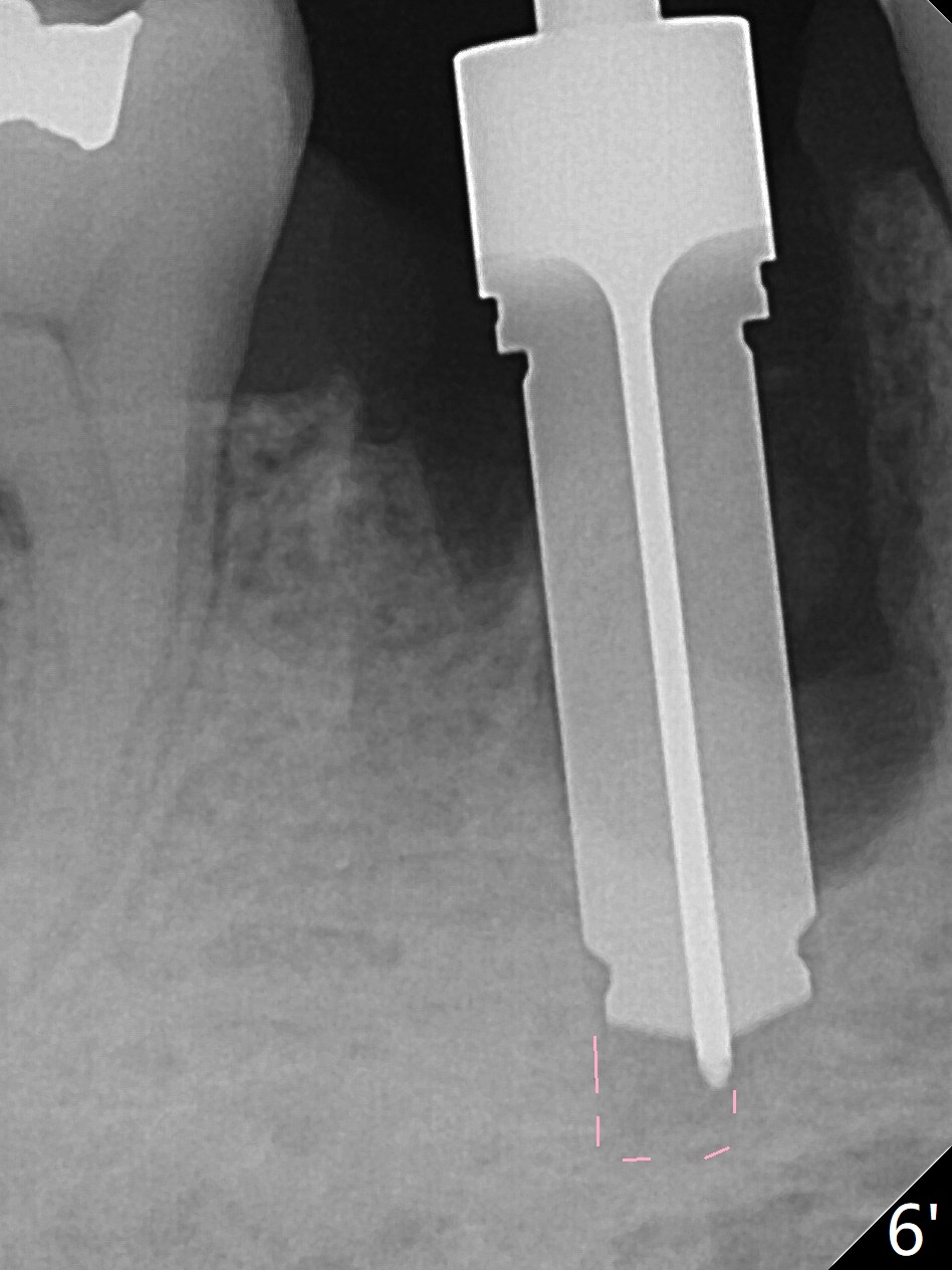
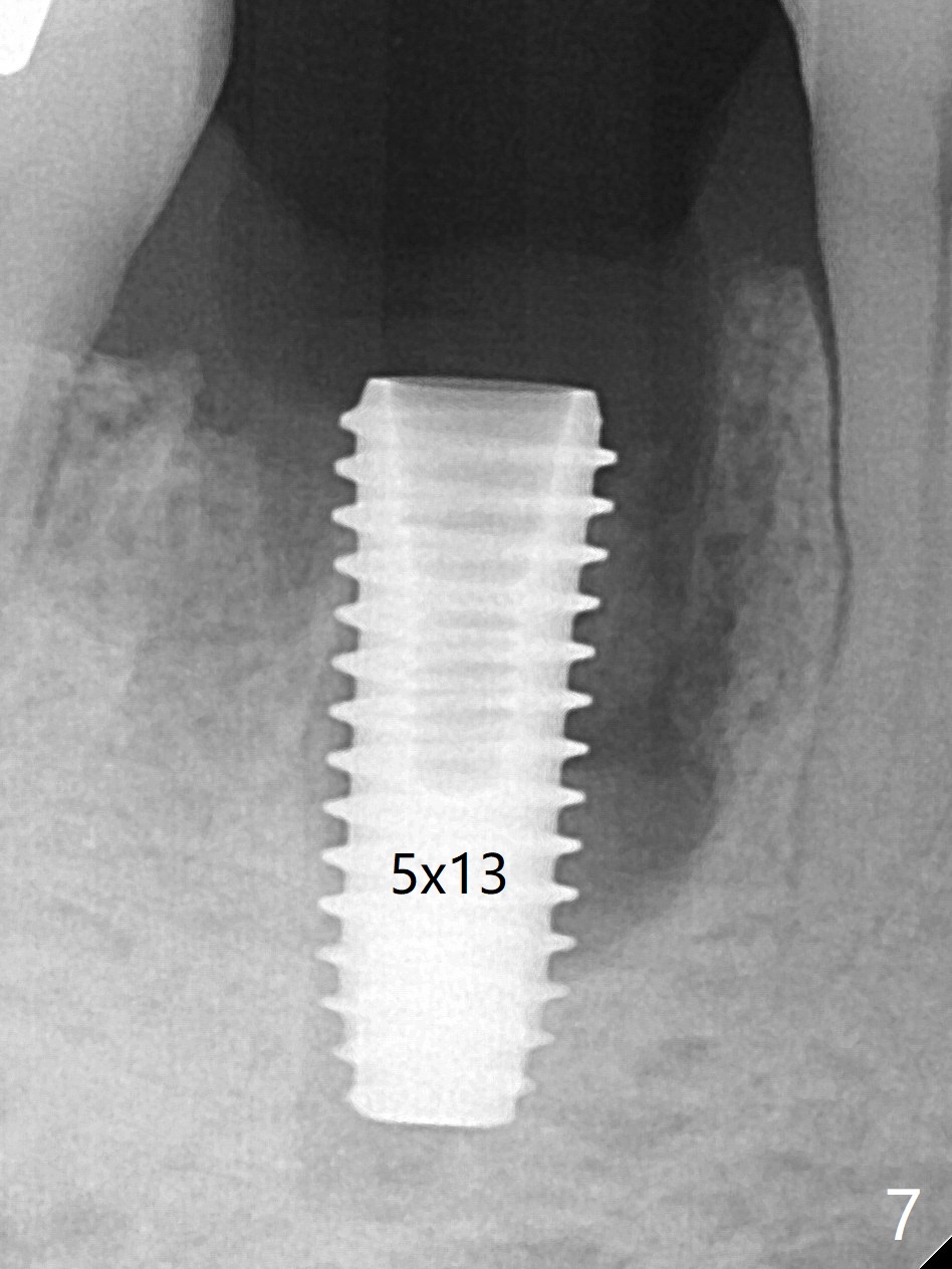
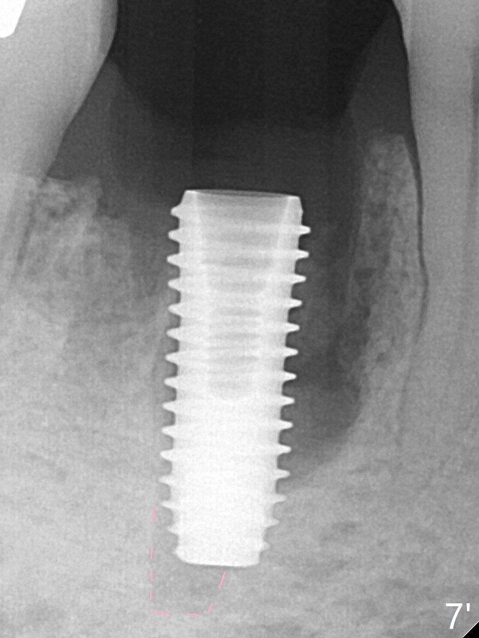
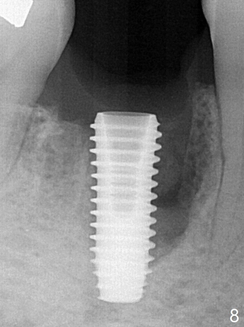
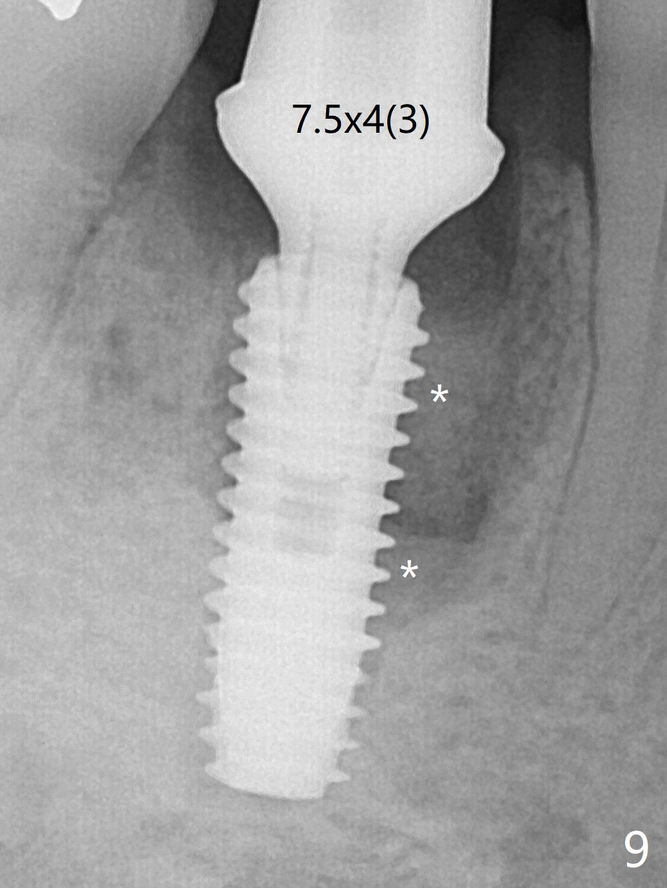
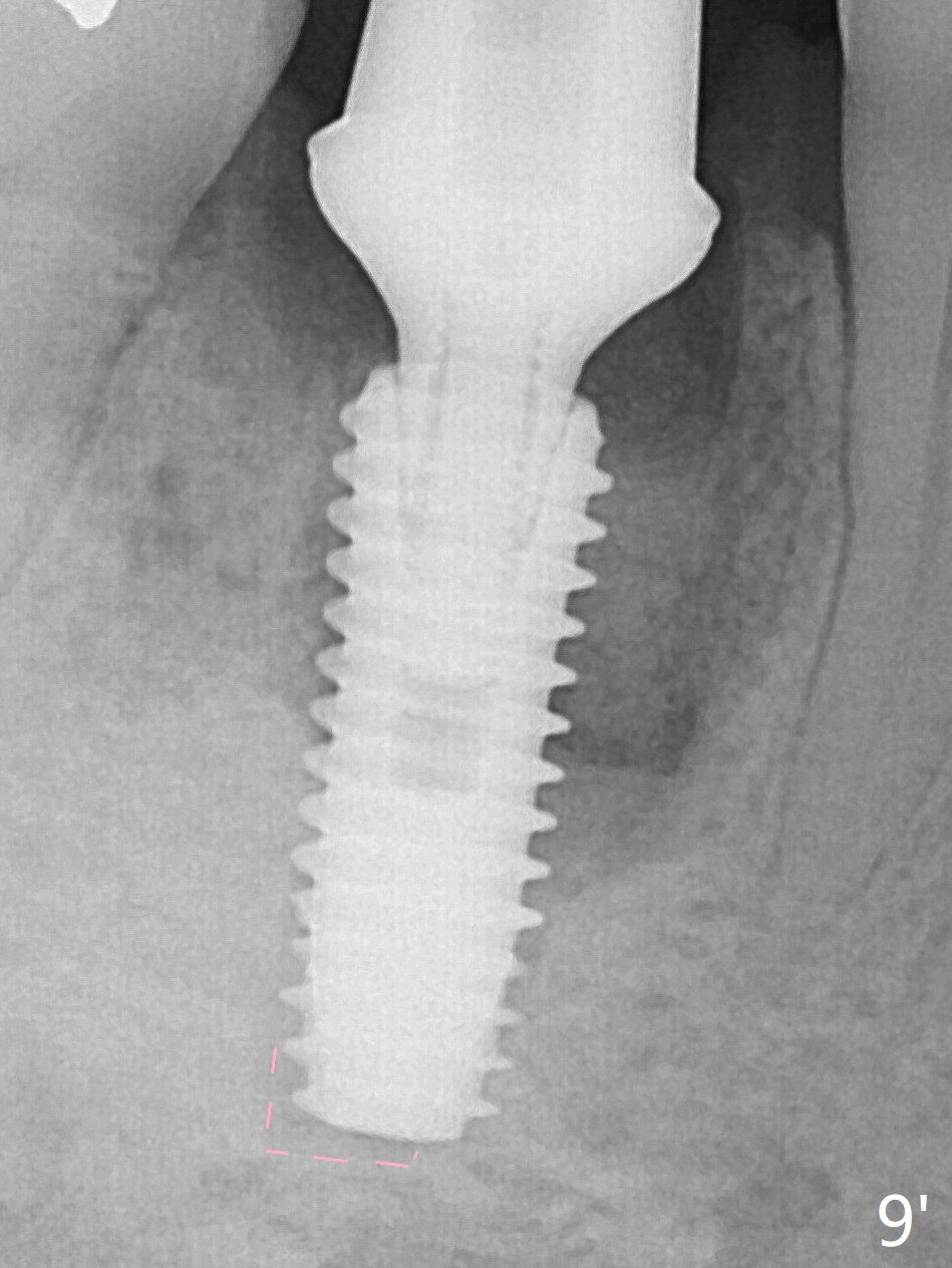
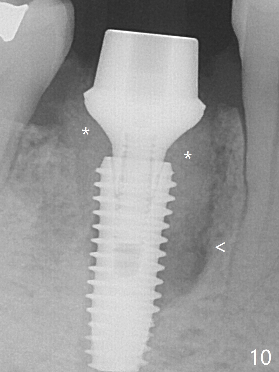
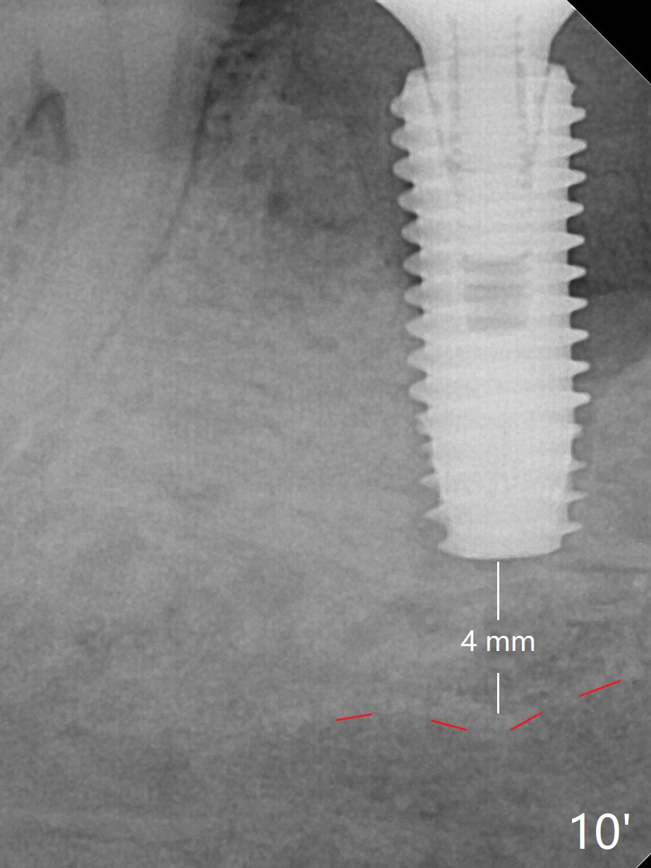
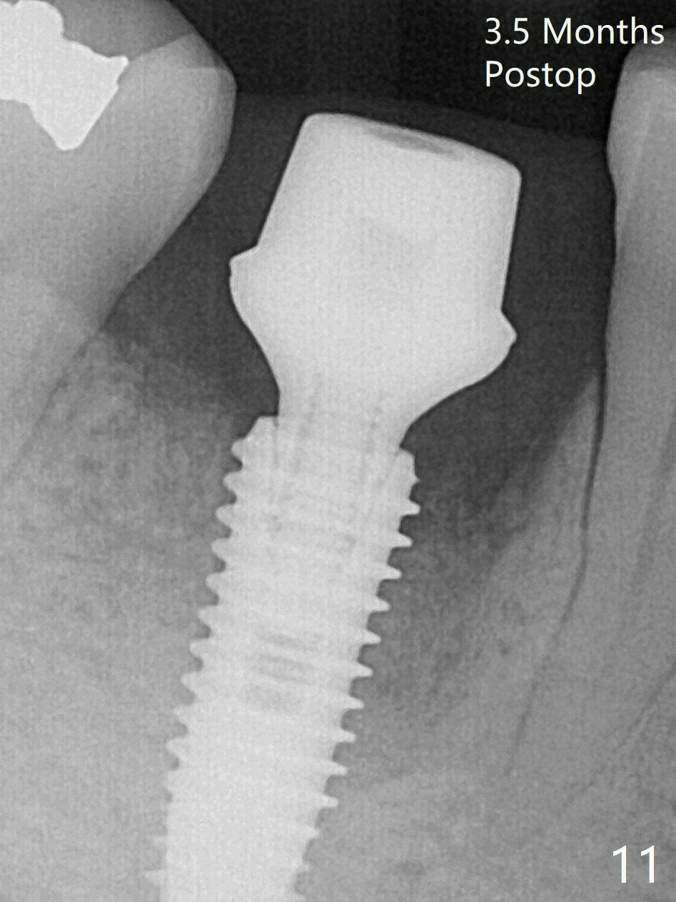
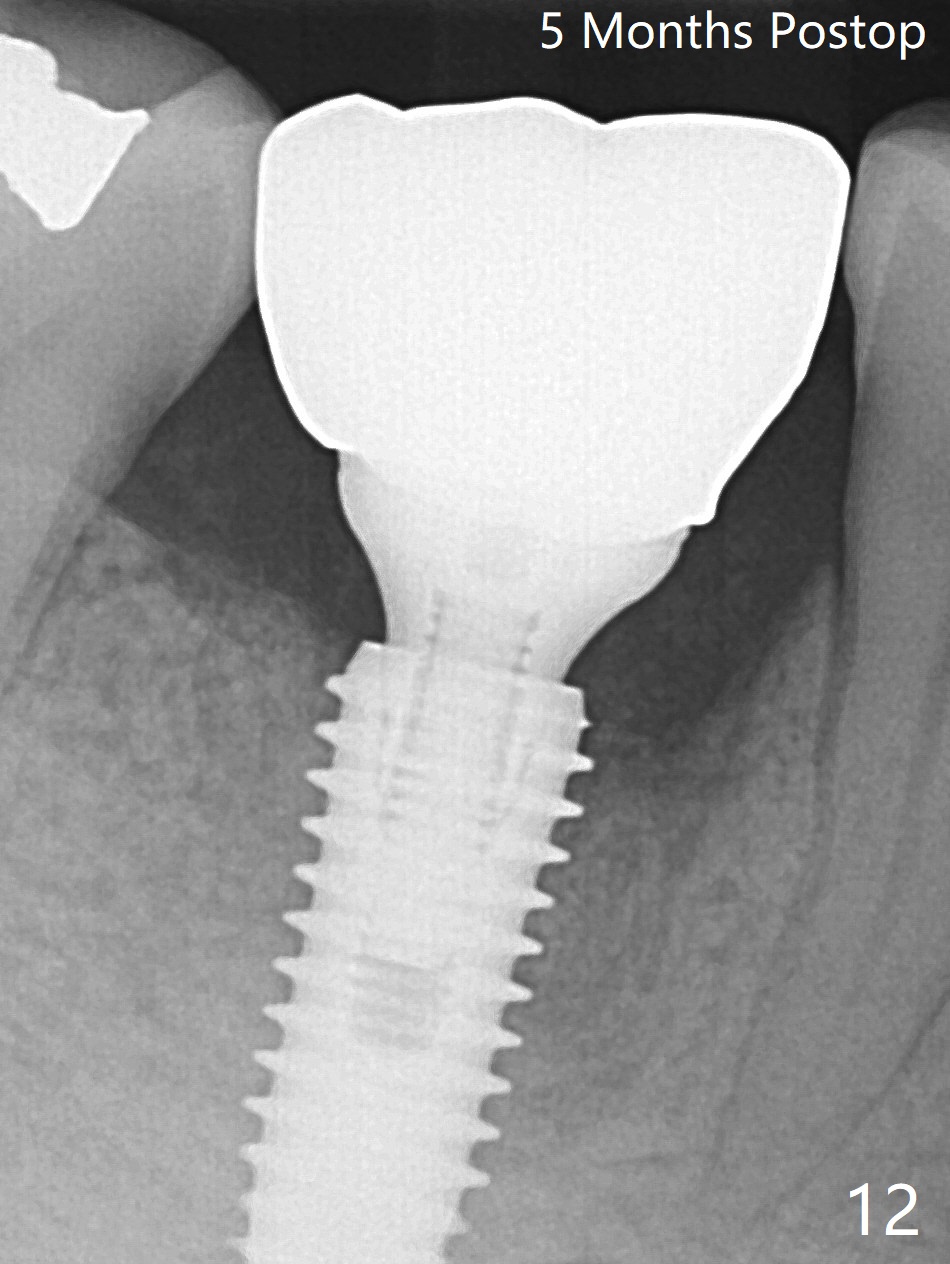
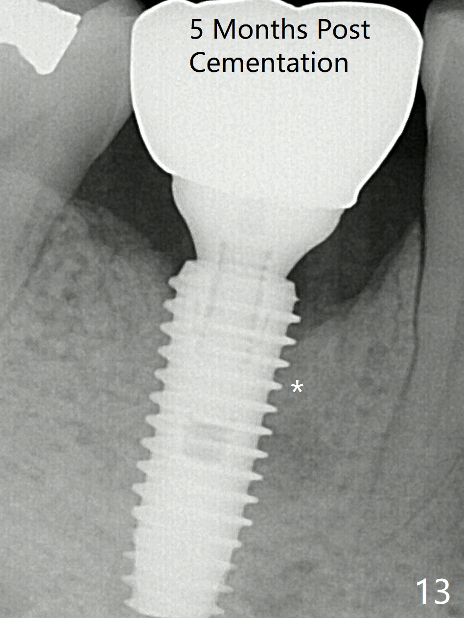
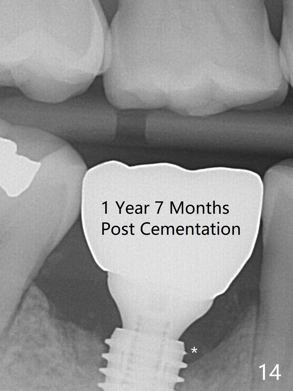
Preop oral Amoxicillin seems to be associated with reduction in the buccal and lingual (Fig.3 arrow) fistulae at #30, but there is mesiobuccal swelling (Fig.1 *) with 7 mm pocket (Fig.2). Osteotomy is initiated in the middle of the septum (Fig.3-5 S). As the osteotomy increases, it shifts mesially (Fig.6 arrow). Guided surgery is able to reduce shifting. A 5x13 mm implant is not seated completely (Fig.7) apparently due to osteotomy shifting. After removal of the bone from the osteotomy distally, the implant remains unseated with lower torque value (Fig.8). Following reuse of the 4.3 mm drill deeper by 1-2 mm, the implant is seated to a satisfactory depth (Fig.9 with increase in torque to 50 Ncm) with placement of Vera Graft (*) and a 7.5x4(3) mm abutment. After a second round of allograft placement (Fig.10 *), the implant is found to be 4 mm from the IAC. At the later stage of osteotomy, the coronal end of the septum is destroyed with loss of osteotomy depth landmark. It is apparent that the soft tissue landmark may be more reliable. The implant threads appear to be covered by the bone graft 3.5 months postop (Fig.11). The abutment is changed to 6.5x5(3) mm one before impression with minor margin prep. The bone density seems to increase 5 months postop, i.e., immediately post cementation (Fig.12) and 10 months postop (5 months post cementation (after retightening abutment), Fig.13 (*)). Periimplantitis develops mesiobuccally, consistent with bone loss 1 year 7 months post cementation (Fig.14 *); the implant seems to have been buccally placed. Bone graft is necessary with PRF or GEM21S if the vein is small and 6-month membrane with a hole around a 7.5x4(4) cemented abutment for easy wound closure. Take 5x5 CM CBCT to determine which wall has defect, buccal or lingual. Check mesial contact. If so, remove the crown, reseat the abutment (possible incomplete seating) and re-impress after bone graft.
Mineralized cortical FDBA. Follow Dr. Stuart Froum 8 step technique for treating peri-implantitis. He has published this technique. I believe it was in Int. J Perio Rest Dent. Also it is in his book. Dr. Sam Lynch 04/21/2020
Return to
Lower
Molar Immediate Implant, Prevent
Molar Periimplantitis (Protocols,
Table),
IBS,
No Deviation
Socket Shield
Prevention
Xin Wei, DDS, PhD, MS 1st edition 10/16/2017, last revision 07/01/2020