|
|
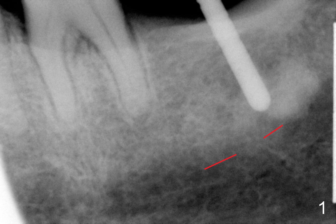
After establishing proper depth of osteotomy in the relatively narrow ridge at #18 (Fig.1 (red dashed line: the superior border of the Inferior Alveolar Canal)), the patient feels pain when the osteotomy is enlarged (Fig.2 with 4.5x8 mm drill in place) in spite of repeated local infiltration. The tooth was extracted 7-8 years ago. It appears that the pain is due to the fact that the osteotomy approaches part of the former periapical radiopaque lesion (Fig.2 *).
Xin Wei, DDS, PhD, MS 1st edition 01/28/2016, last revision 07/11/2018