|
|
|
|
|
|
|
|
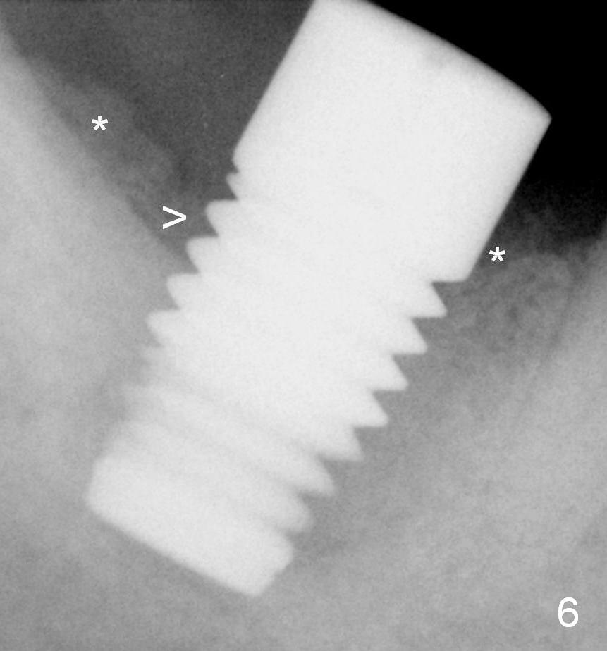
X-ray 2 months postop shows bone graft (Fig.6 *) deposited near implant threads (>).
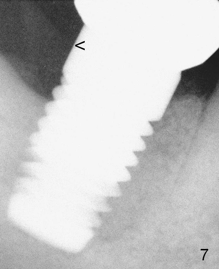
The definitive restoration is delivered 3 months postop. Fig.7 is taken 1 month post cementation; the crown margin is supragingival (<: gingival margin), which is easy for cement removal.
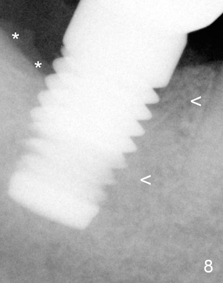
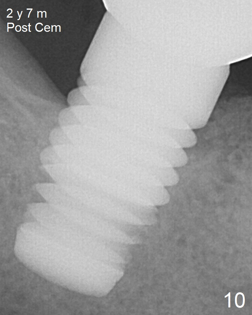
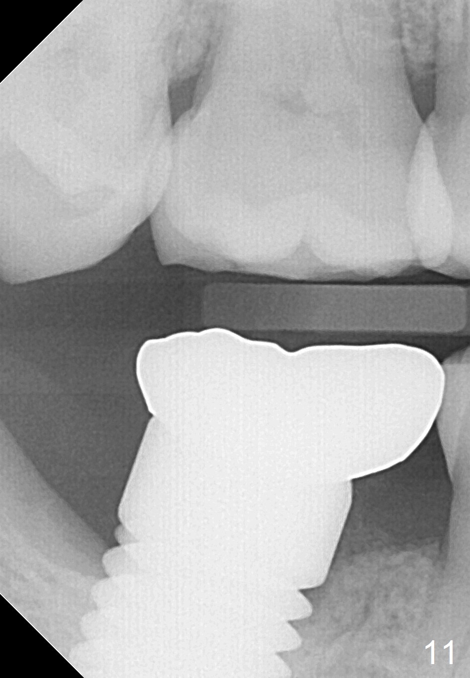
Seven months post cementation (10 months postop), the bone graft remains distal to the implant (Fig.8 *), while bone density mesial to the implant continues to increase (<). There is no bone loss 2 years 7 months post cementation (Fig.10,11).
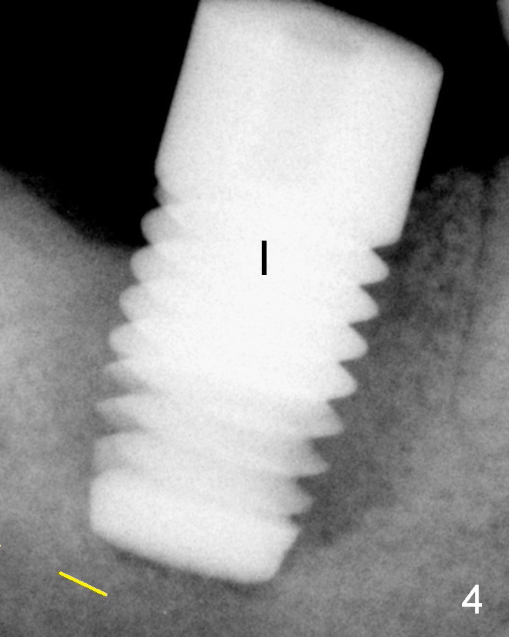
Bone density around the implant increases as time passes (Fig.4,6,7,8,10,11).
Implant Forms Osteotomy Course 1 2 Last Next
Xin Wei, DDS, PhD, MS 1st edition 11/16/2014, last revision 12/22/2018