|
|
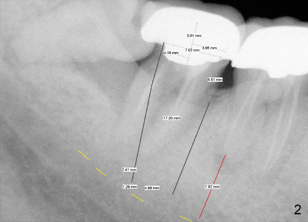
Fig.2 (preop PA) is placed deep enough to show both the upper (yellow dashed line) and lower borders of the inferior alveolar canal (IAC). The distal apex of the 1st molar is a reliable landmark for the position of the upper border of the IAC when a PA does not show the whole IAC.
There is severe bone loss between the 2nd and 3rd molars. The distal margin of #31 crown is open.
Return to Bone Injection
Xin Wei, DDS, PhD, MS 1st edition 09/30/2014, last revision 09/30/2014