.jpg)
|
|
|
|
|
|
|
|
|
|
High Torque Value Due to Cortical Bone
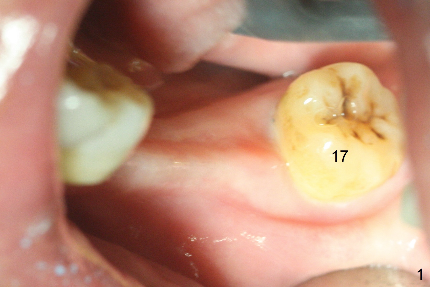
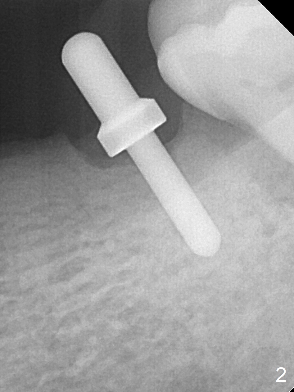
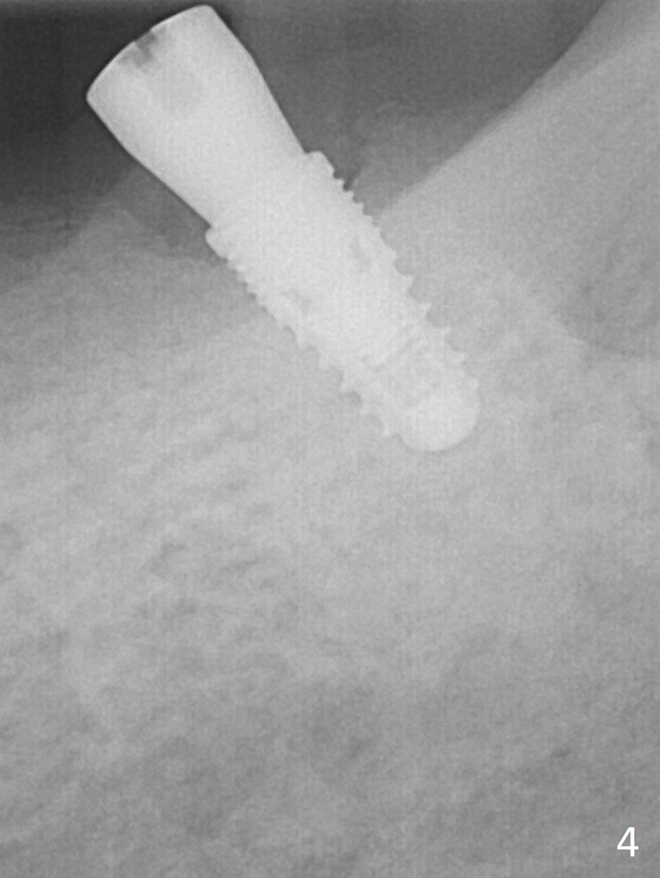
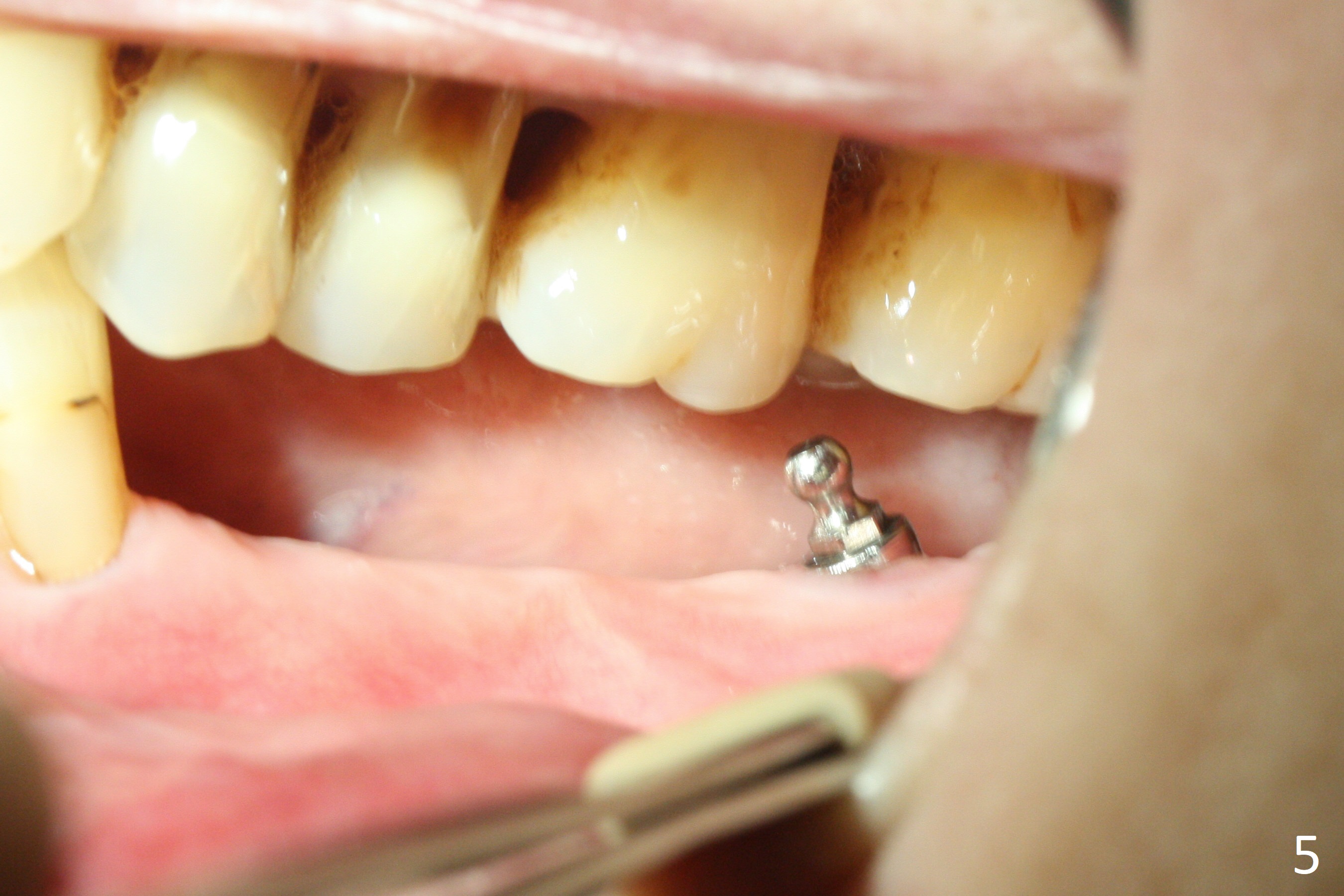
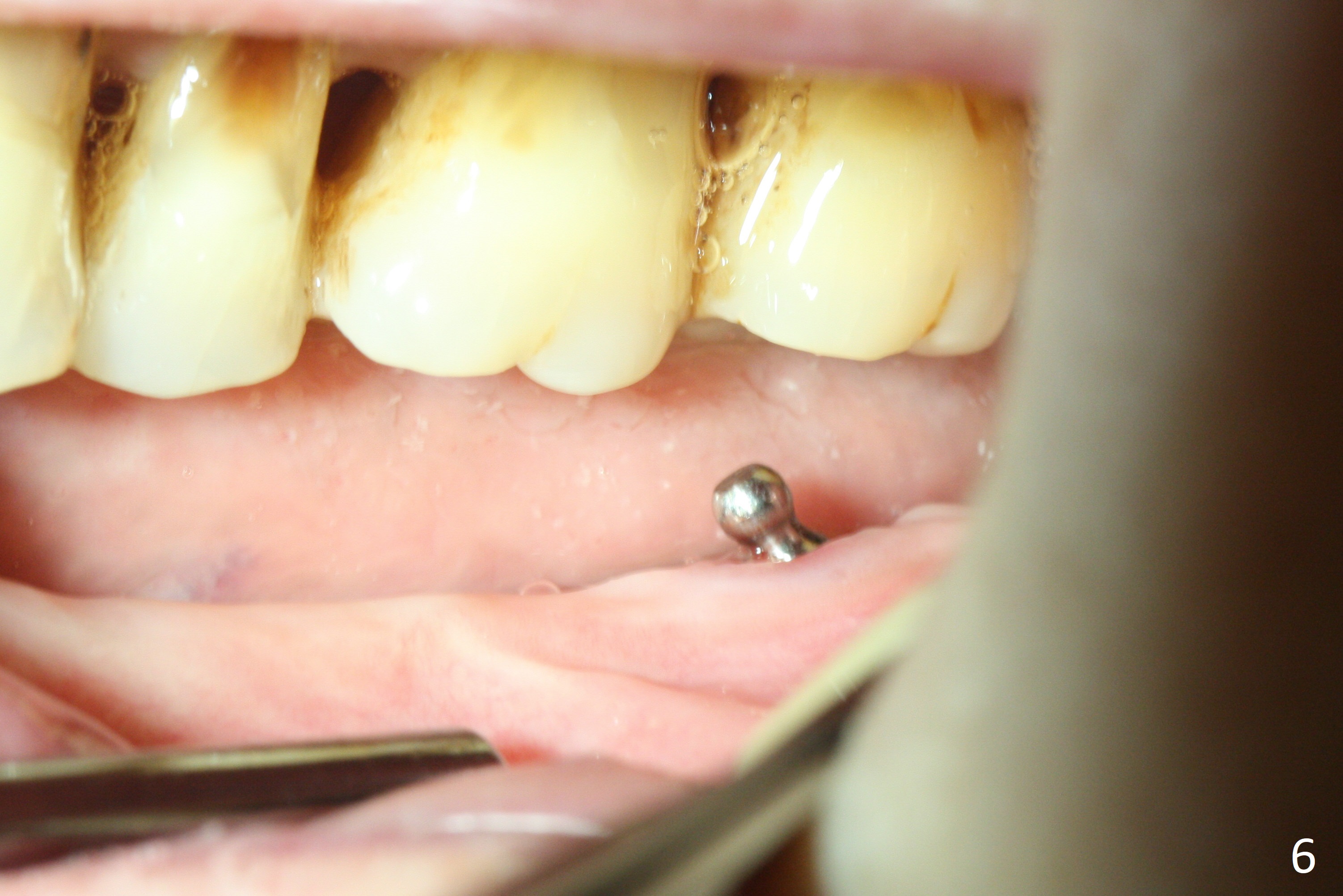
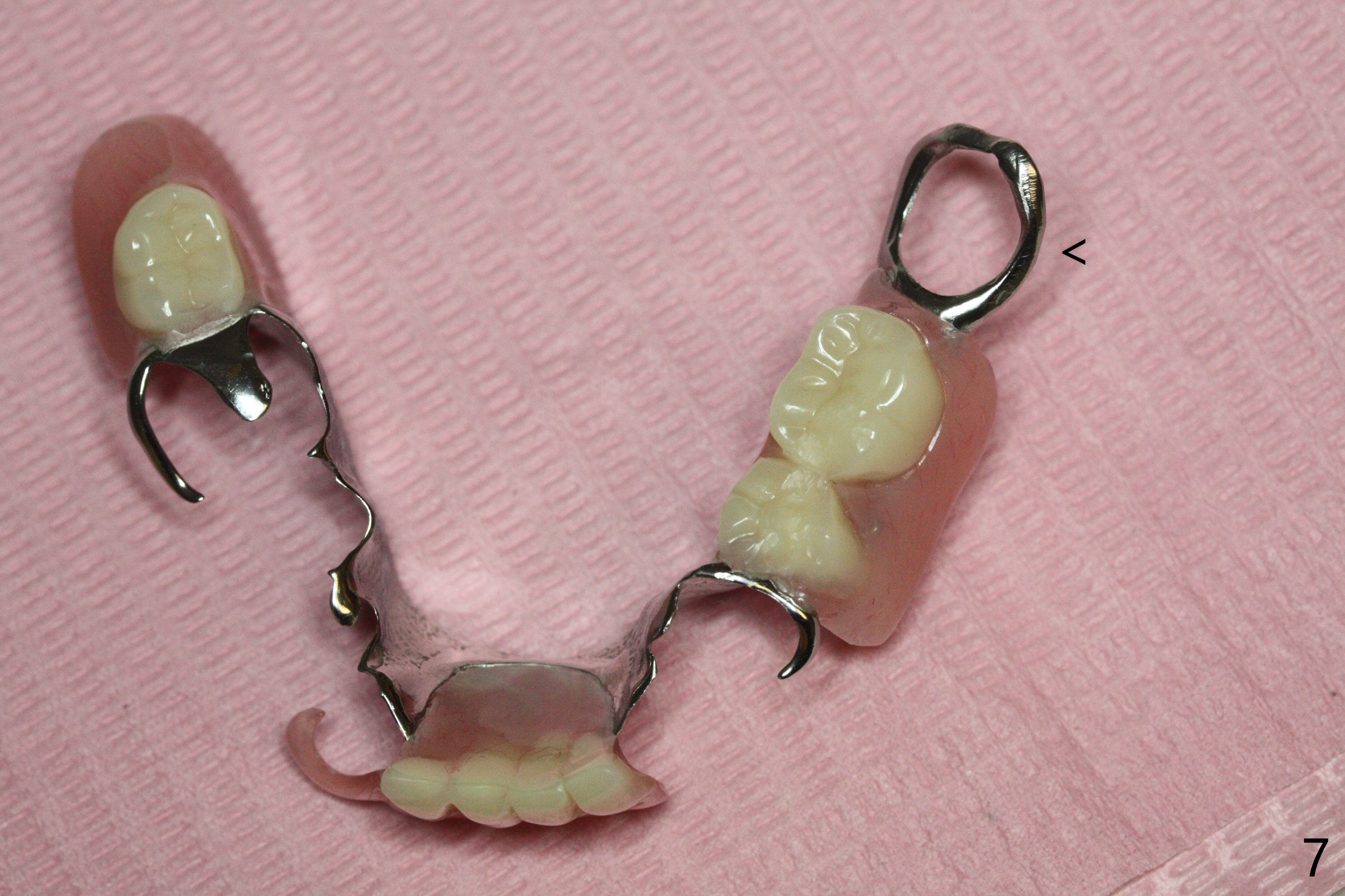
Preop photo confirms the lower left posterior pointed ridge (Fig.1). After ridge reduction and 2 mm drill for 8 mm, a parallel pin is inserted (Fig.2). A 3.8x8 mm SM implant is placed with >50 Ncm (Fig.3). The coronal threads (buccodistal) is covered with VeraGraft after placement of 4.1x5(3) mm healing abutment. The tooth #17 is not planned for extraction while the implant is osteointegrated. The tooth appears to be too loose and is extracted ~ 1.5 months postop. The implant seems to be osteointegrated 3 months postop (Fig.4). The healing abutment is changed to ball abutments with 4 mm (Fig.5) and 2 mm (Fig.6) cuffs. In fact the latter stays. What is the special device at the site of #18 (ring, Fig.7 <)?
Return to
Lower
Molar Immediate Implant, IBS
2nd Case of Ball Abutment for RPD
Xin Wei, DDS, PhD, MS 1st edition 08/18/2017, last revision 03/01/2018