|
|
|
|
|
|
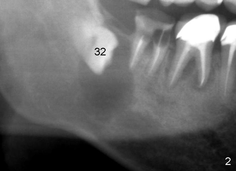
Fig.2: Preop Pan
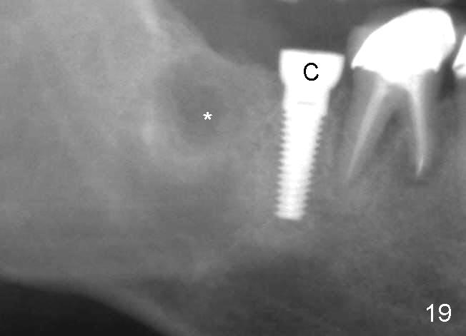
Fig.19: Postop 6 months. The cystic lesion appears to have been decreased concentrically (*). C: healing cuff.
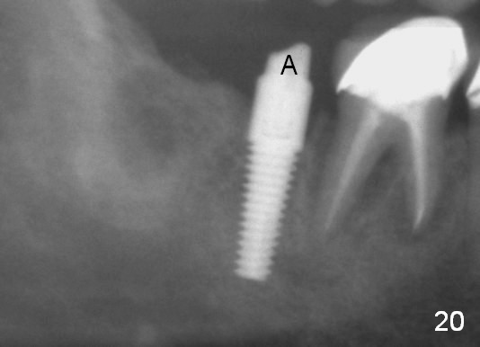
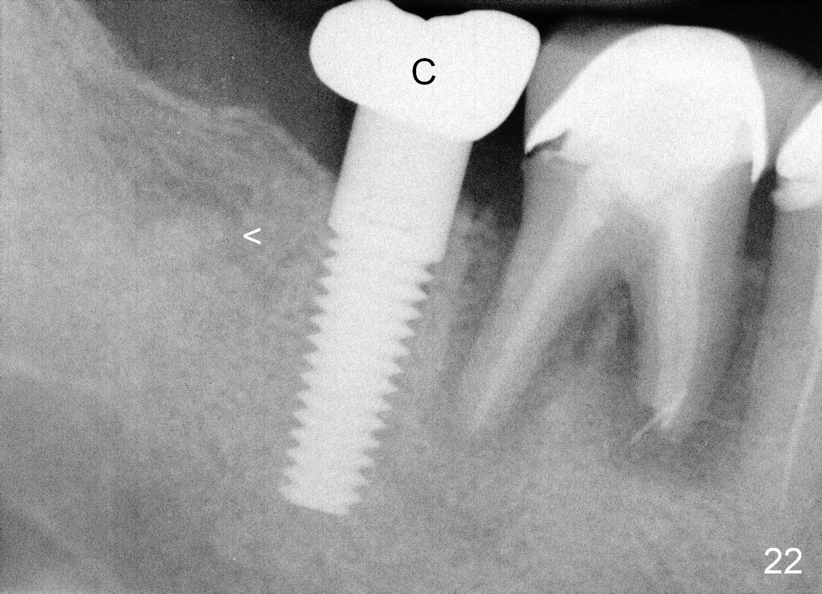
The bone density in the former cyst area continues to increase 14 (Fig.20 A: abutment) and 32 (Fig.22) months postop. Small piece of bone graft (<) is being extruded distobuccal to the #31 crown (C) asymptomatically.
Return to Dentigerous Cyst Last Next Preop Pan 图三
Xin Wei, DDS, PhD, MS 1st edition 03/28/2013, last revision 09/18/2017