|
|
|
|
|
||
|
|
|
|
|||
|
|
|
|
|||
|
|
|
|
|||
|
|
|
|
|||
Removal of Two Mesiodens Followed by Immediate Implants and Provisionals
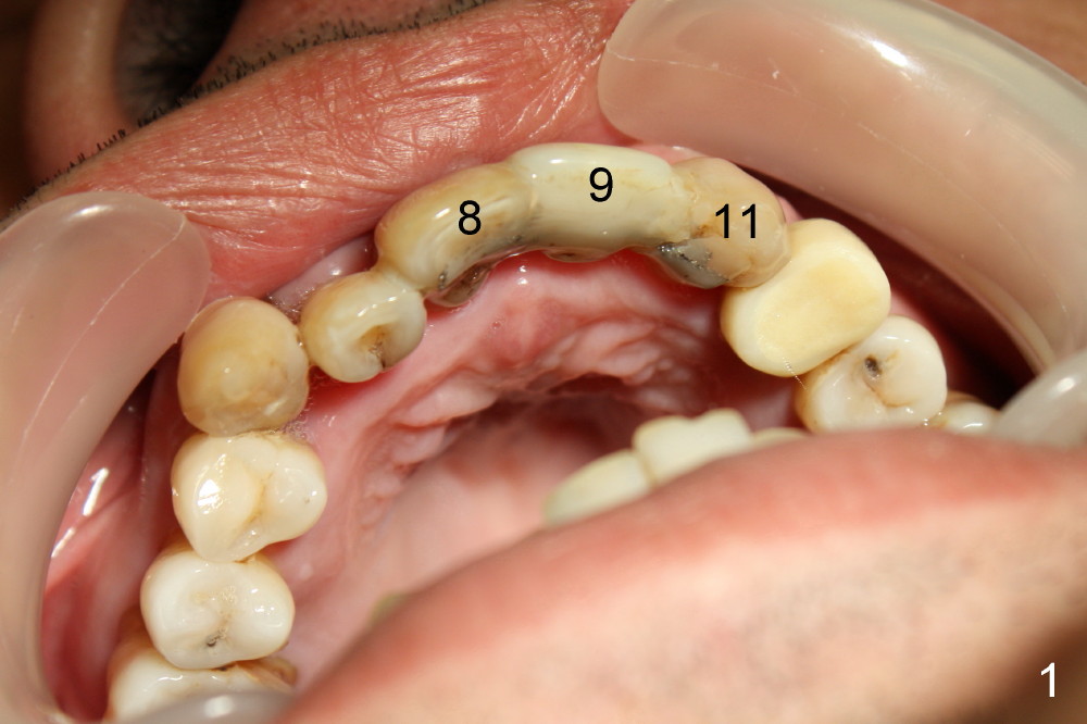
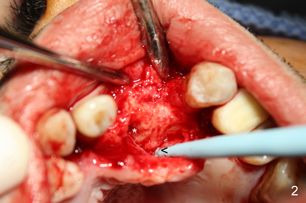
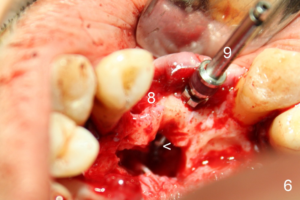
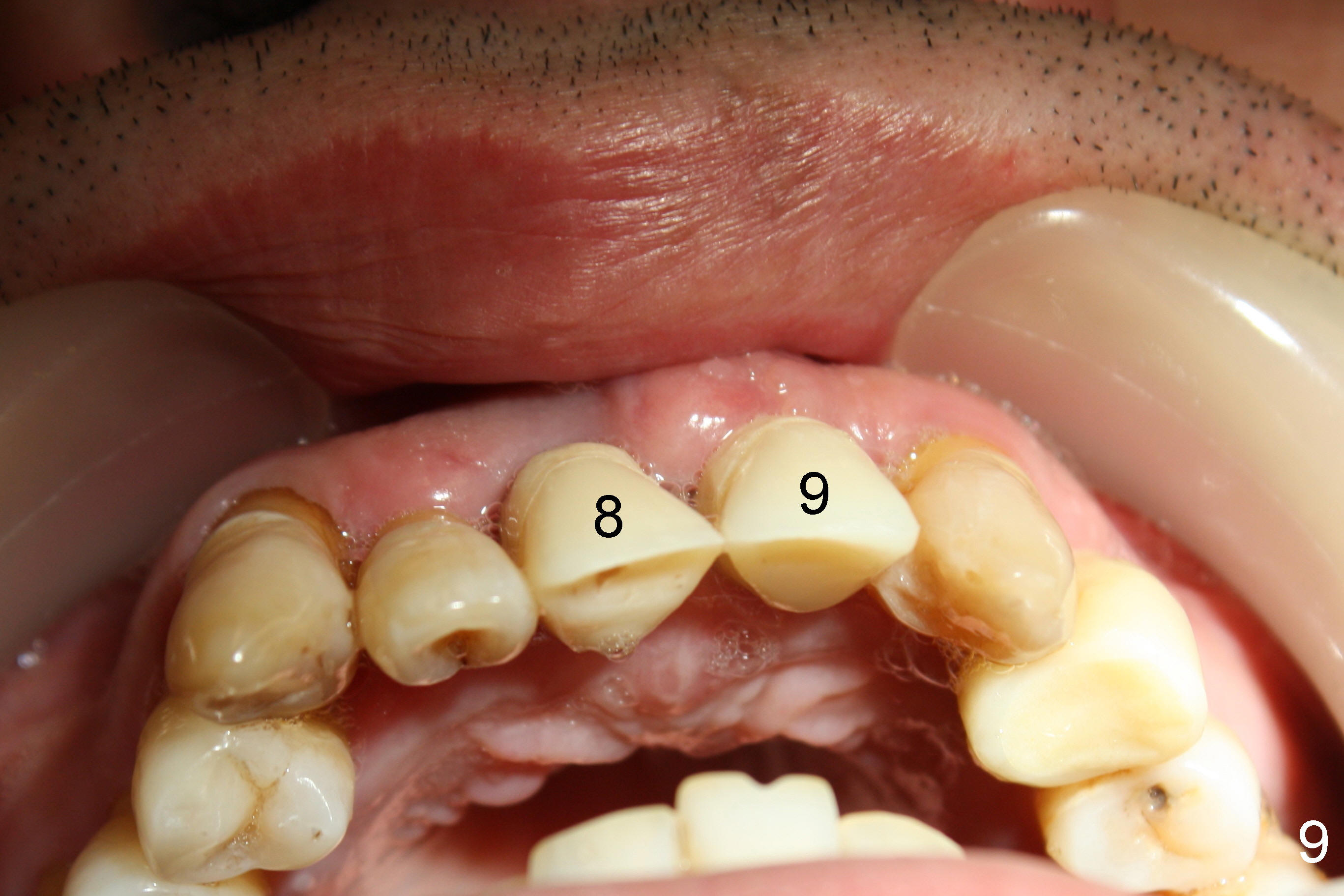
CBCT shows that the two mesiodens, esp. left one, are close to the palatal plate. After removal of the peridontally compromised tooth #8 and the pontic #9 (as compared to Fig.1), the palatal flap is raised to expose the left mesiodens (Fig.2 <; *: incisal foramen).
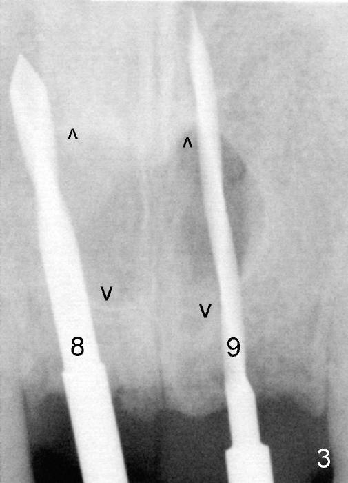
Following extraction of the two mesiodens, osteotomy starts with pilot drills (Fig.3: 8,9). As planned, these pilot drills go through two of the cortical plates of the mesiodens' sockets (arrowheads).
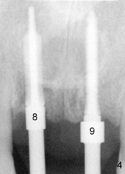
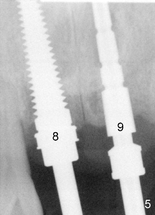
Trajectory is being corrected while osteotomy is increasing with reamers (Fig.4: 3 and 2.5 mm). When a 4.5x20 mm tap is inserted at the site of #8 (Fig.5), it is stable. A 3.5x20 mm tapered drill is at the site of #9 for parallelism. When the tap is removed from the osteotomy of #8, the middle portion of the 3.5x20 mm drill at the site of #9 is seen to pass the socket of the left mesiodens (Fig.6: <).
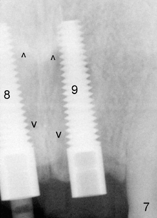
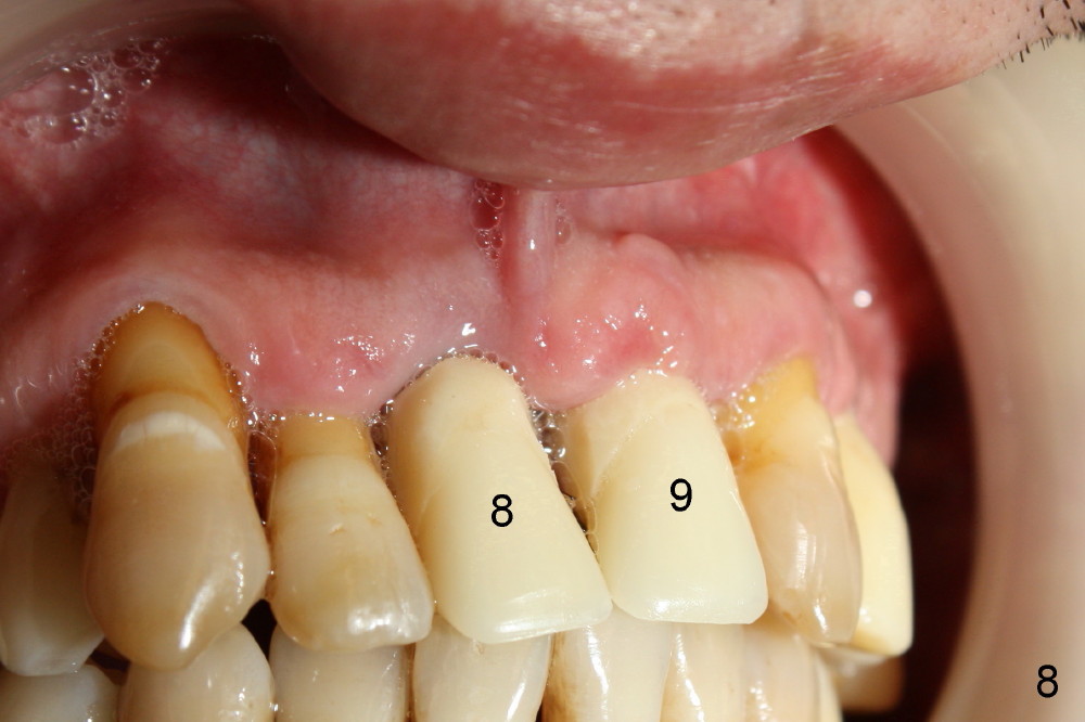
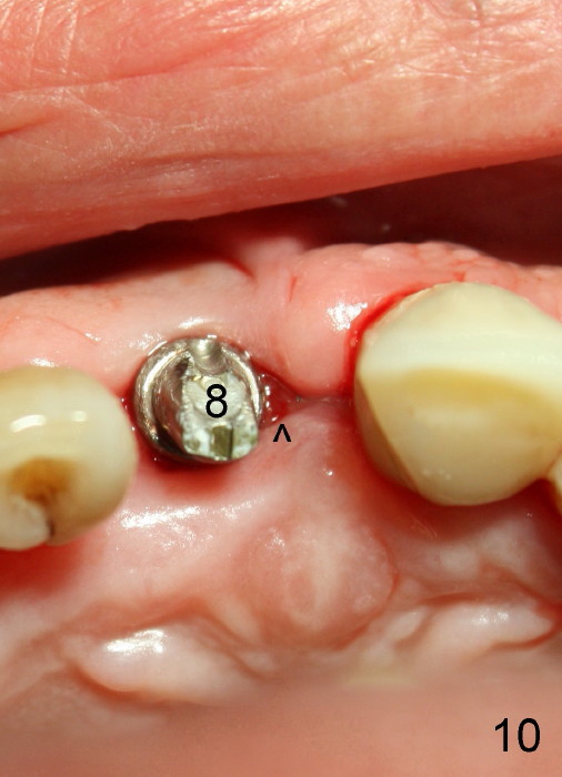
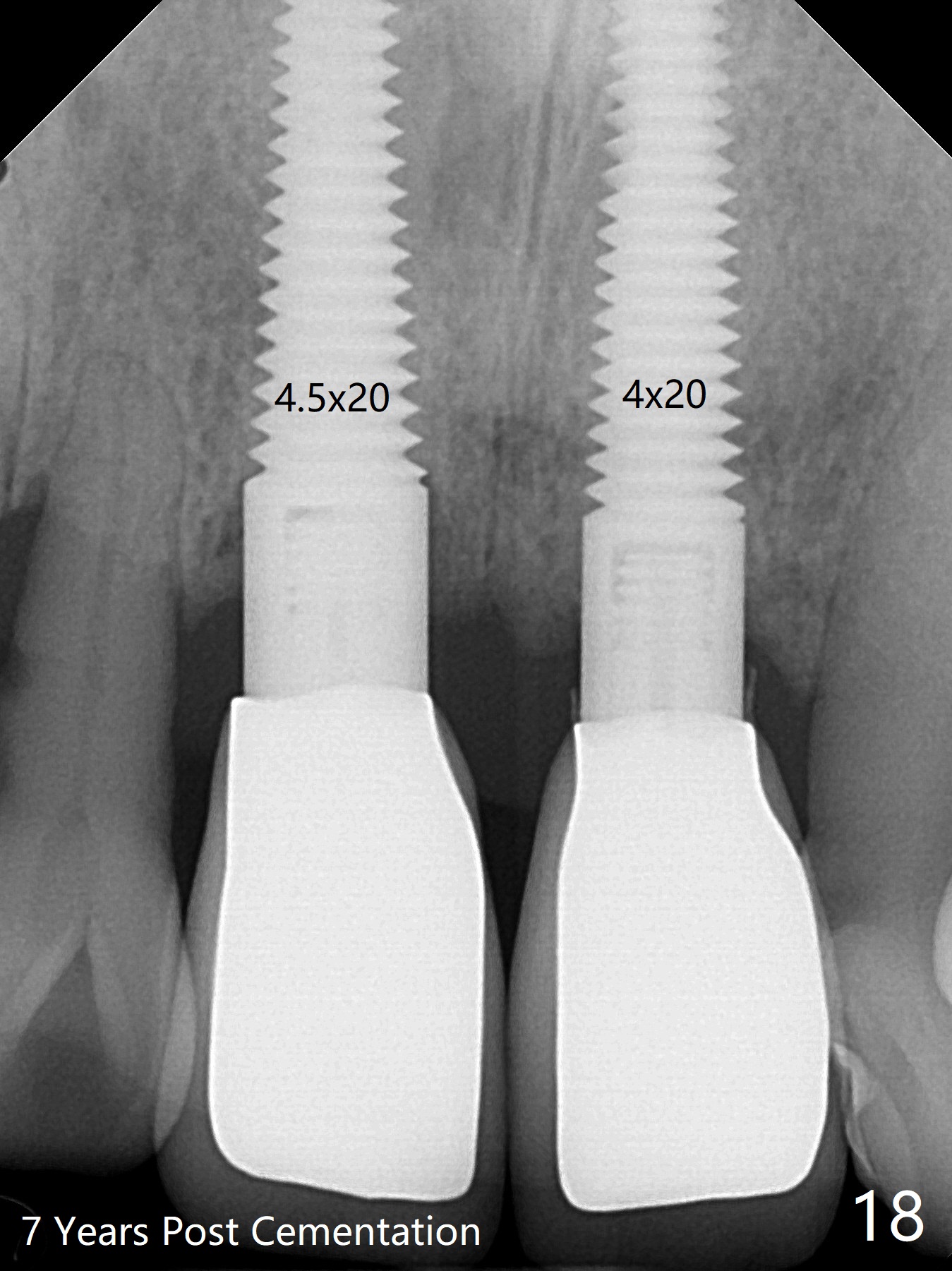
The insertion torque for the implants (4.5x20 mm and 4x20 mm) are 40 and 60 Ncm for #8 and 9, respectively (Fig.7). The stability is probably due to the fact that these two implants are engaged into the two cortical plates of the sockets (arrowheads). Allograft is placed in the mesiodens sockets. Immediate provisionals are fabricated. Fig.8,9 show the provisionals 18 days postop. One of the provisionals (#8) is dislodged 5 weeks postop. Before recementation of the provisional (Fig.10), healthy granulation tissue is found next to the implant (^).
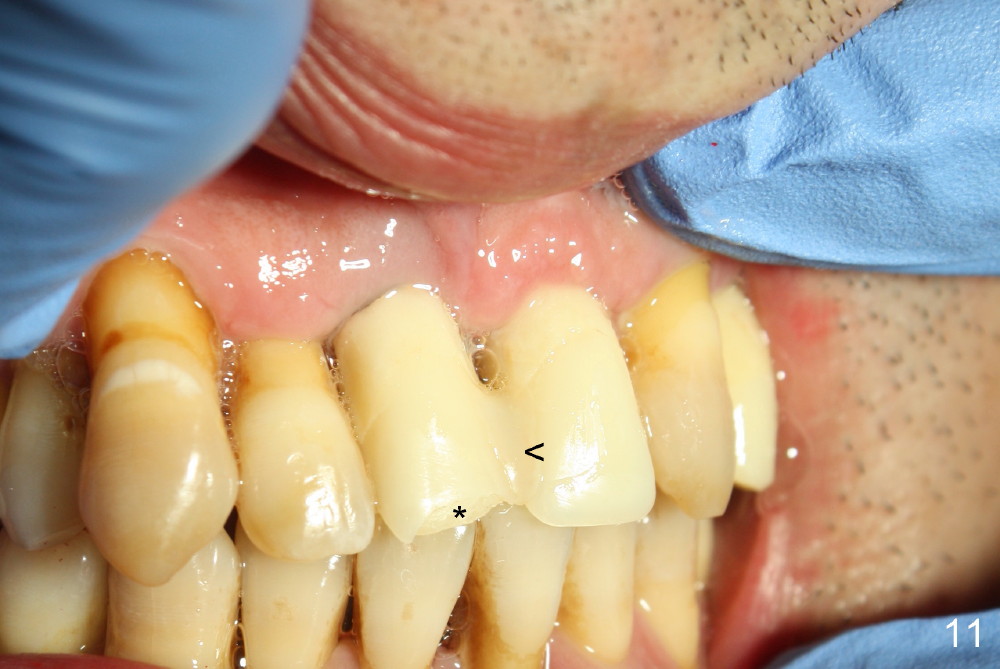
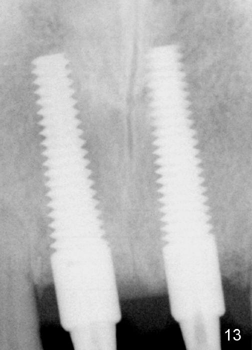
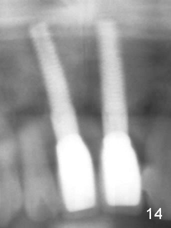
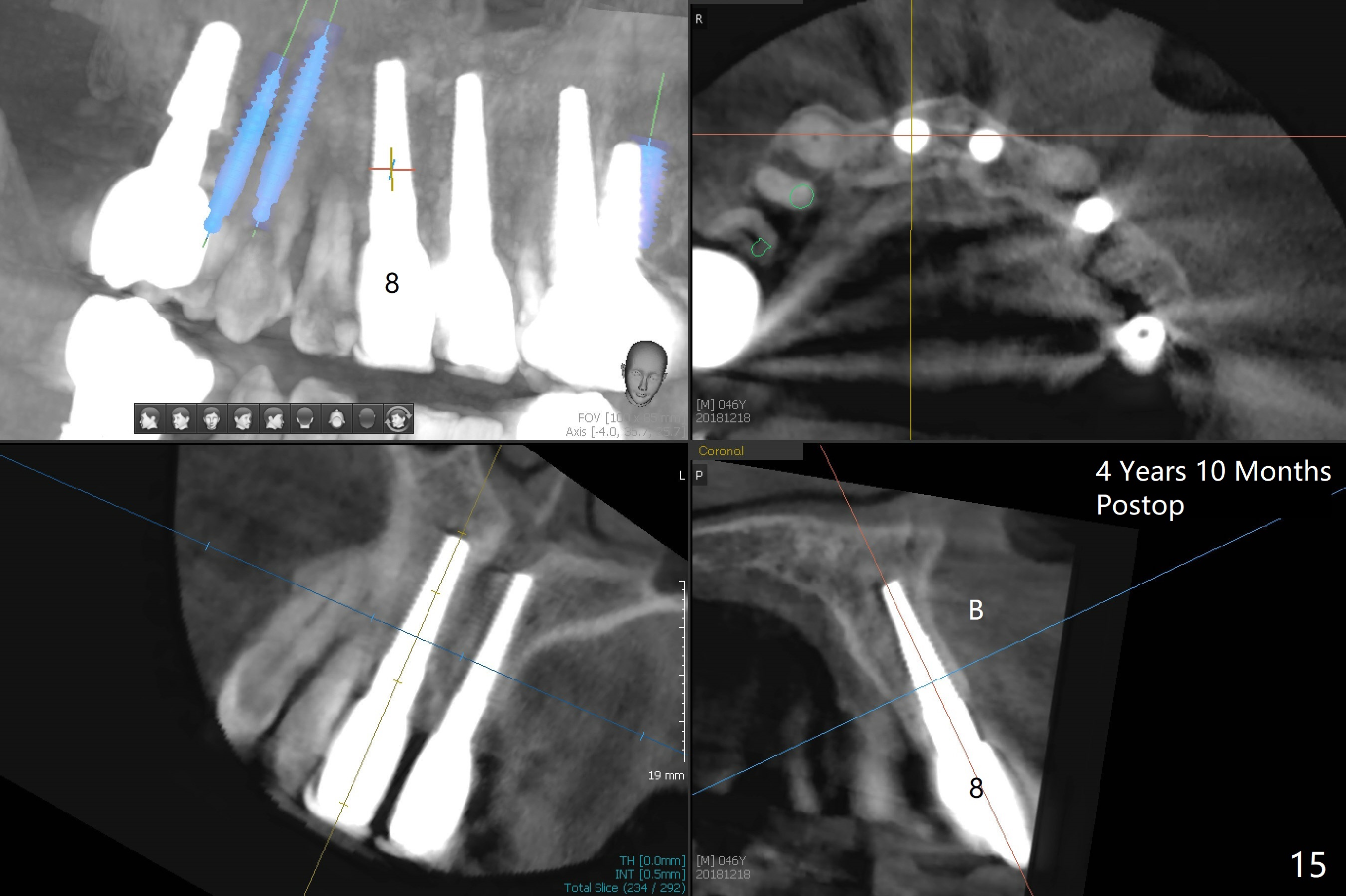
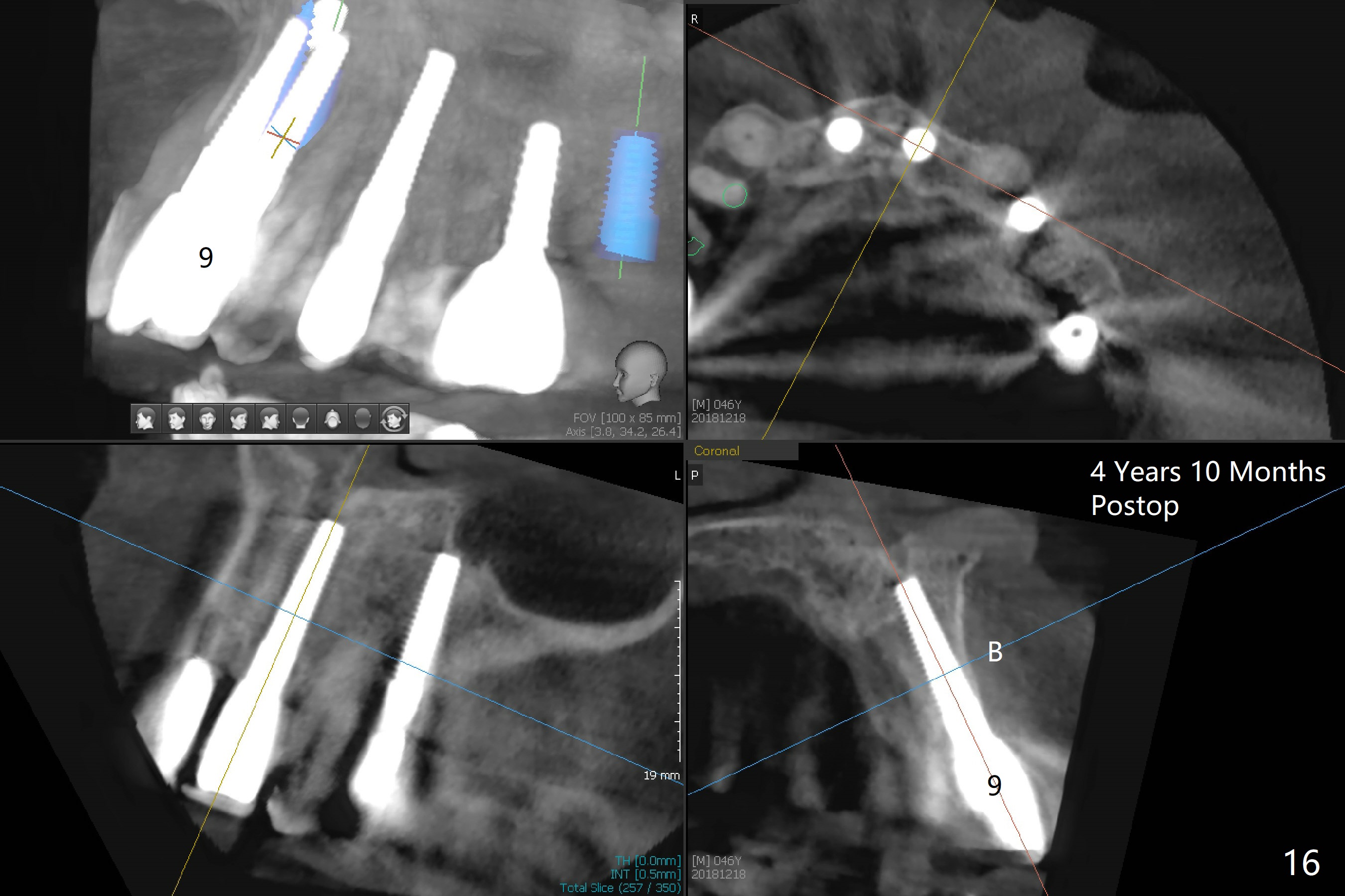
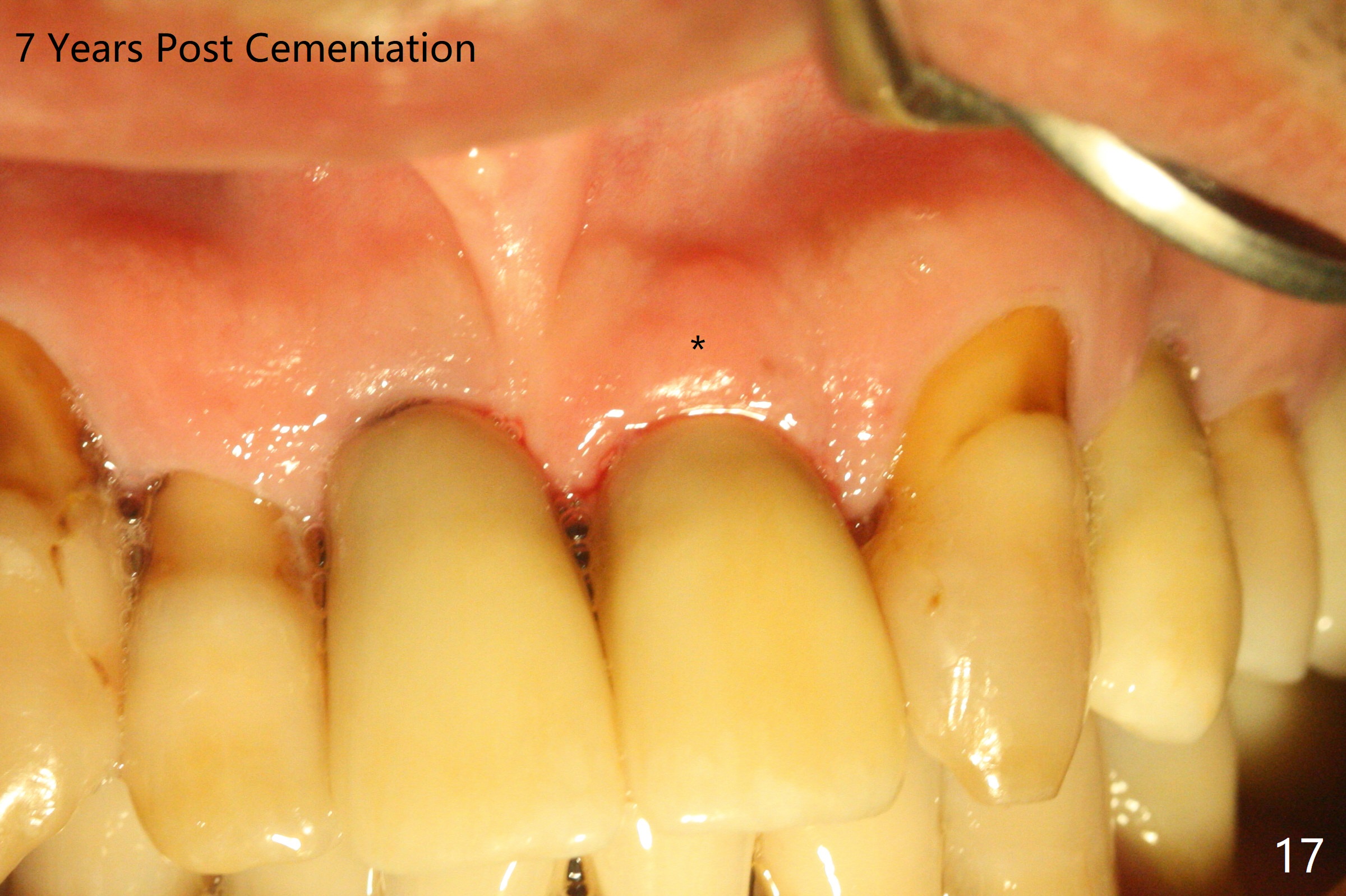
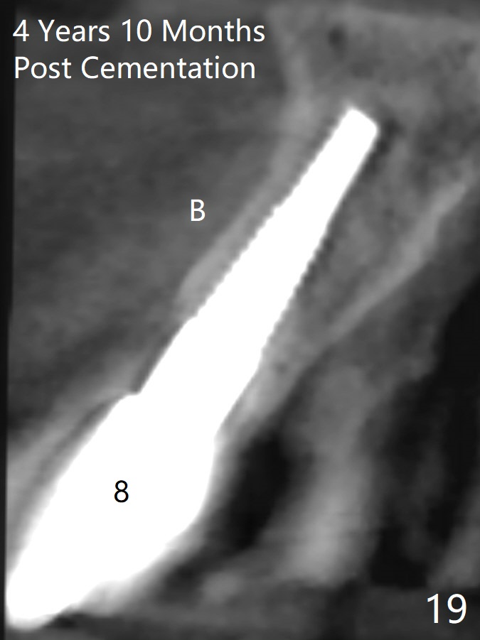
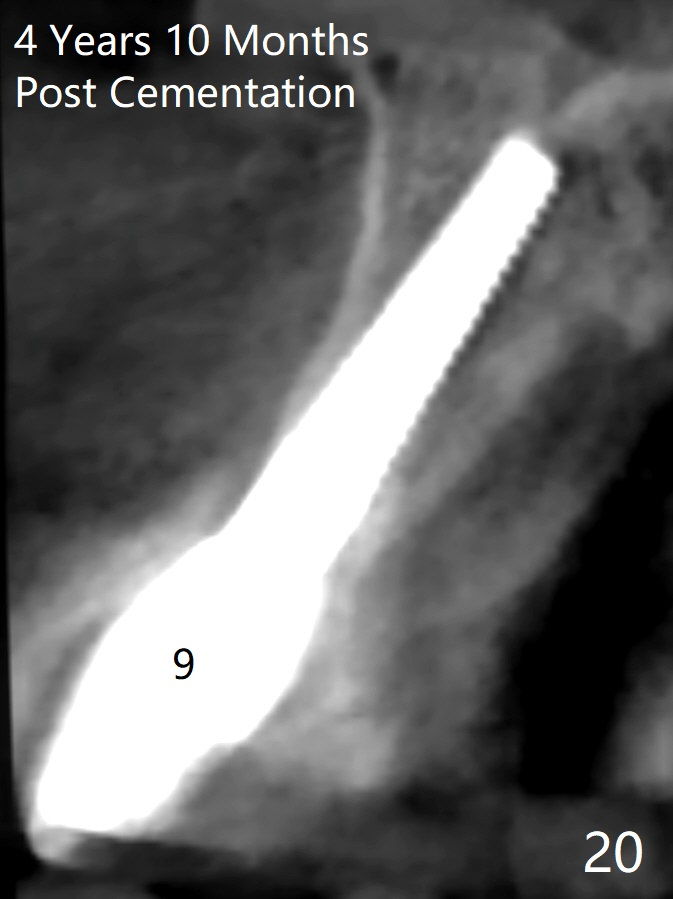
These two anterior implants remain stable 2 months 20 days postop, since one of the immediate provisionals has incisal chip (Fig.11 *) while both of them are splinted (<) to increase retention. There is no complain about paresthesia around the incisal papilla due to separation of the nasopalatine nerve. Mesiodens sockets disappear 7 months postop (Fig.13). There is no bone loss 2 years 9 months postop (Fig.14 panoramus) or 4 years 10 months postop (Fig.15,16 CT). The buccal gingiva is reddish and swollen with bleed on probing and history of pain 7 years post cementation (Fig.17). There is no bone loss in PA (Fig.18). The buccal plate at #9 is thinner than that at #8 4 years 10 months post cementation (Fig.19,20).
Design and Preparation,
Upper Incisor Immediate
Implant,
Dr. Wu Protect
Graft
Xin Wei, DDS, PhD, MS 1st edition 02/03/2014, last revision 07/03/2021