|
|
|
|
|
|
|
|
|
|
|
|
IS or IBS for Thin Bone? M
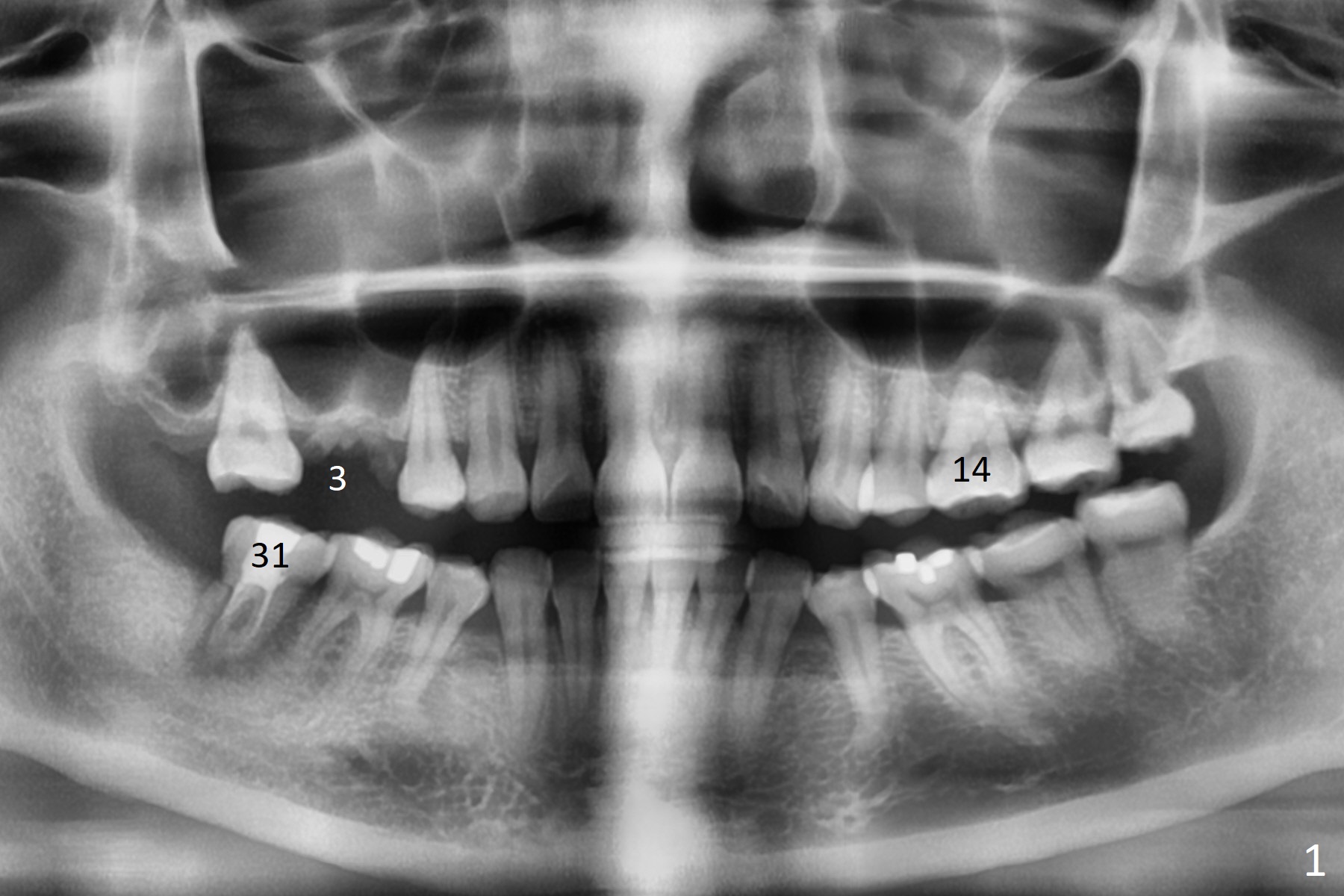
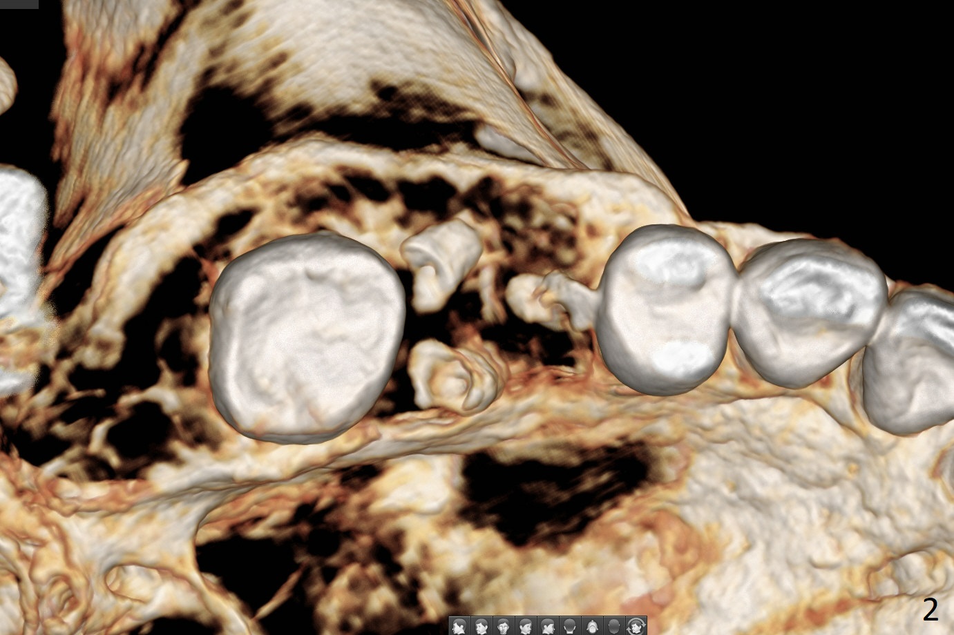
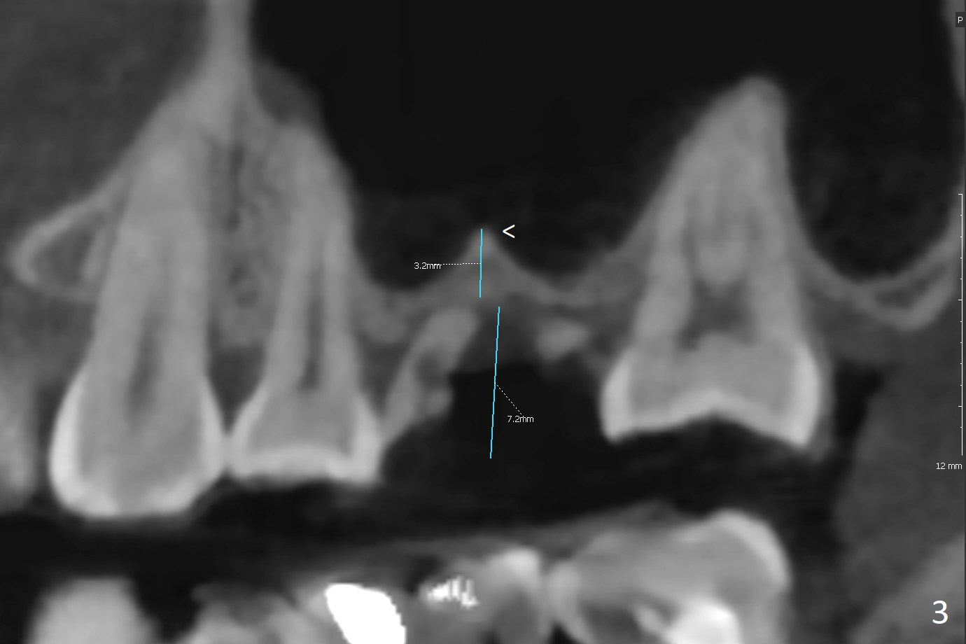
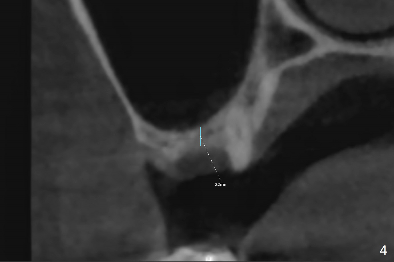
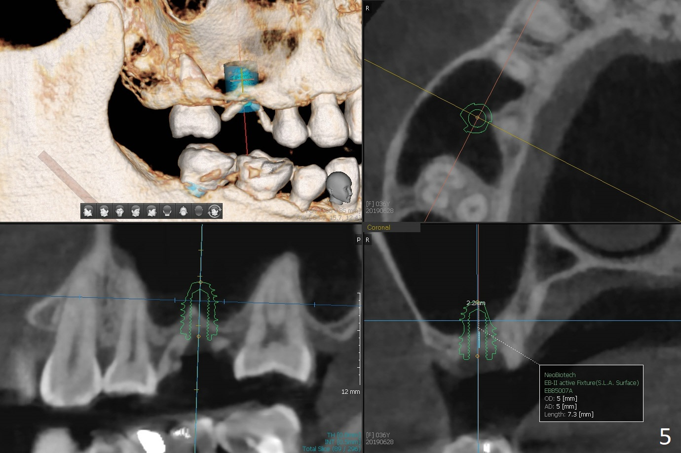
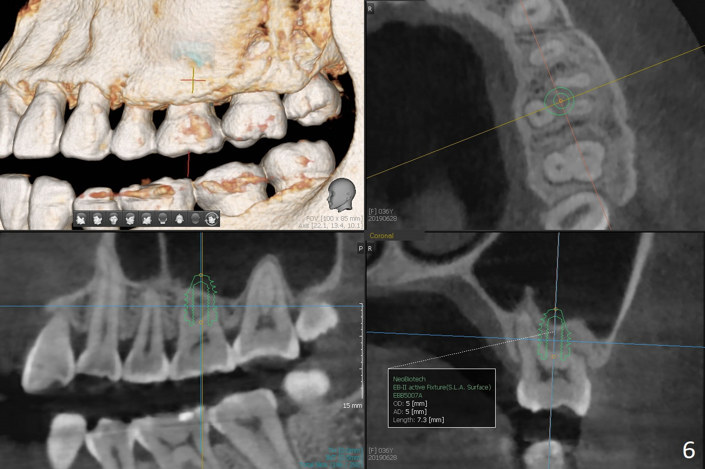
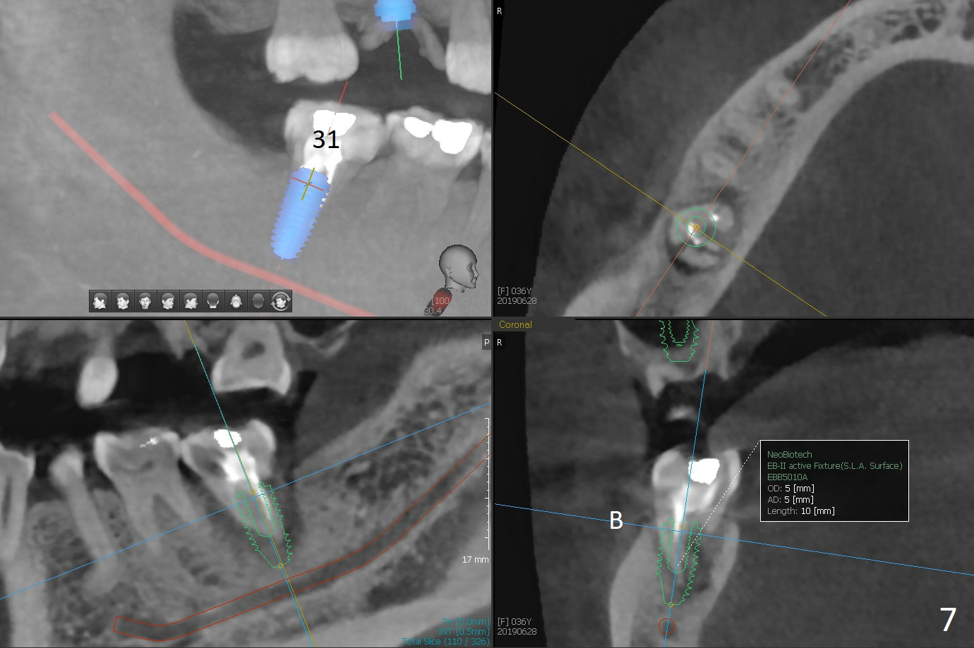
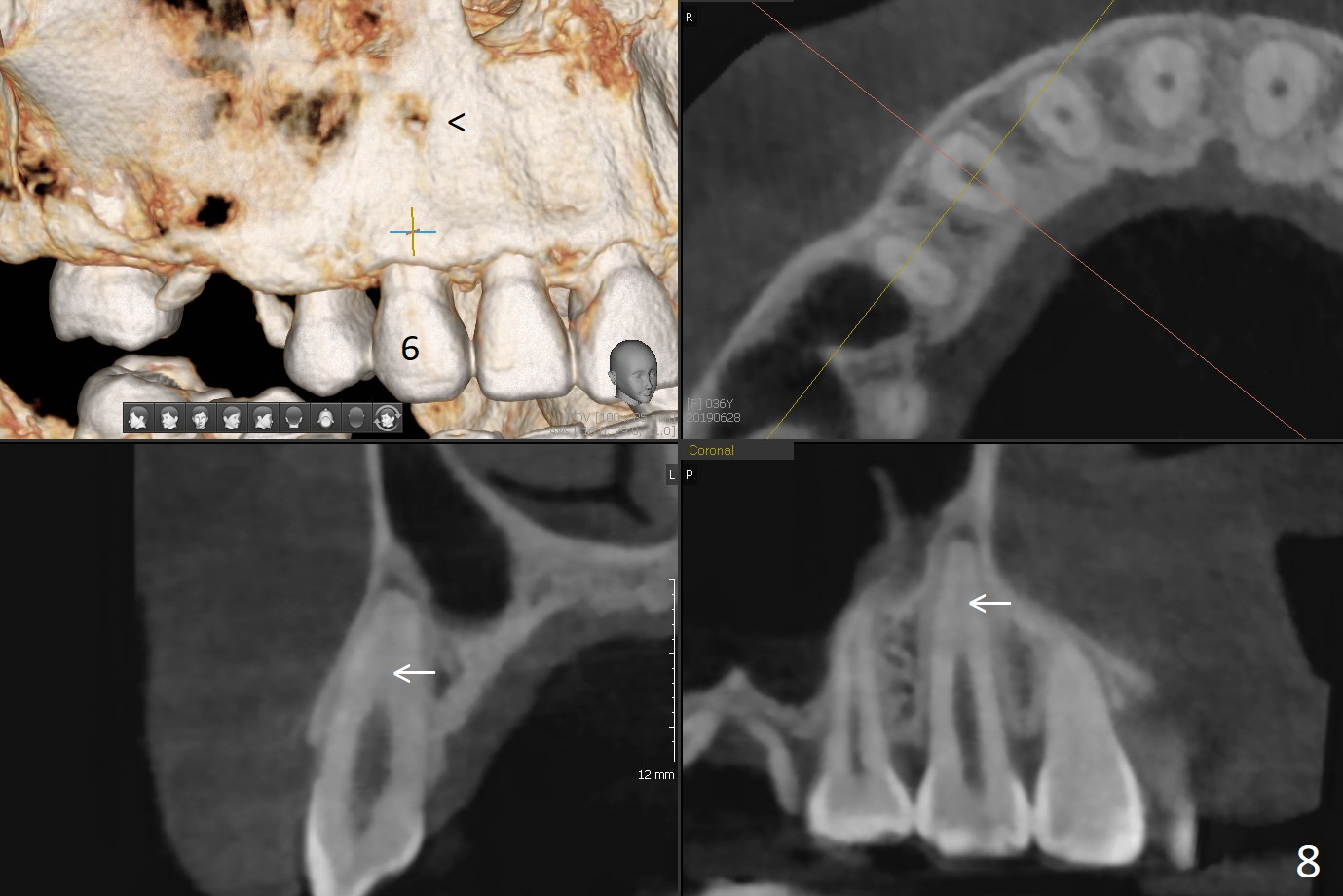
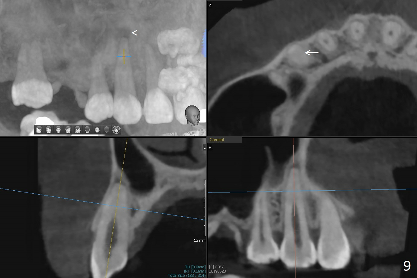
A 36-year-old woman is nervous about dentistry. She will take Valium by herselft before surgery (Fig.1). The tooth #3 has 3 residual roots (Fig.2 (CBCT 3 D occlusal view)). The bone is 2-3 mm thick (Fig.3,4 (sagittal, coronal sections)). A 5x7.3 mm implant will be placed with IS guide (Fig.5 (12 mm offset)). Since IBS implants are able to achieve amazing stability in the thin bone, prepare the shortest 4-5 mm in diameter dummy implants (IS (better surface treatment) and IBS) after sinus lift using UF Guided Sinus Lift Approach Kit (surgery). With intact tooth structure at #14, the bone height is not much (Fig.6), congenital (genetic) in nature. The infection at #31 is more severe (Fig.7). Extraction will be the 2nd in order. Because of limited bone, it is better to do bone graft first. The patient is concerned about the discolored upper right canine, which should be associated with orthodontics 20 years ago (take photos). The apical canal is obliterated (Fig.8,9 arrow) with periapical radiolucency (arrowhead). In fact the bone at #31 is so little that the tooth will be extracted for socket preservation.
Return to
Upper
Lower
Molar Immediate Implant,
Prevent Molar Periimplantitis (Protocols,
Table),
Trajectory II,
No Antibiotic
Xin Wei, DDS, PhD, MS 1st edition
06/28/2019, last revision
01/25/2020