,%205x7.3,%206x5.jpg)
%20mm,%20later%203-4%20more%20turn.JPG)
|
|
|
|
|
|
|
|
|
|
|
|
|
|
|
|
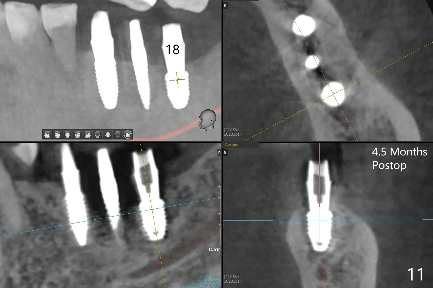
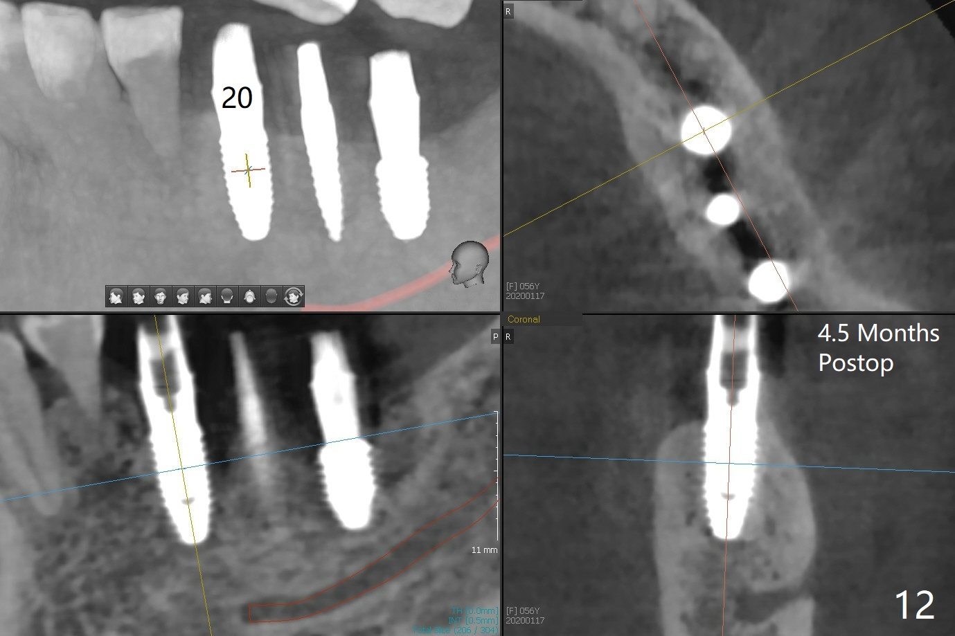
Healing Abutment for Small Socket I
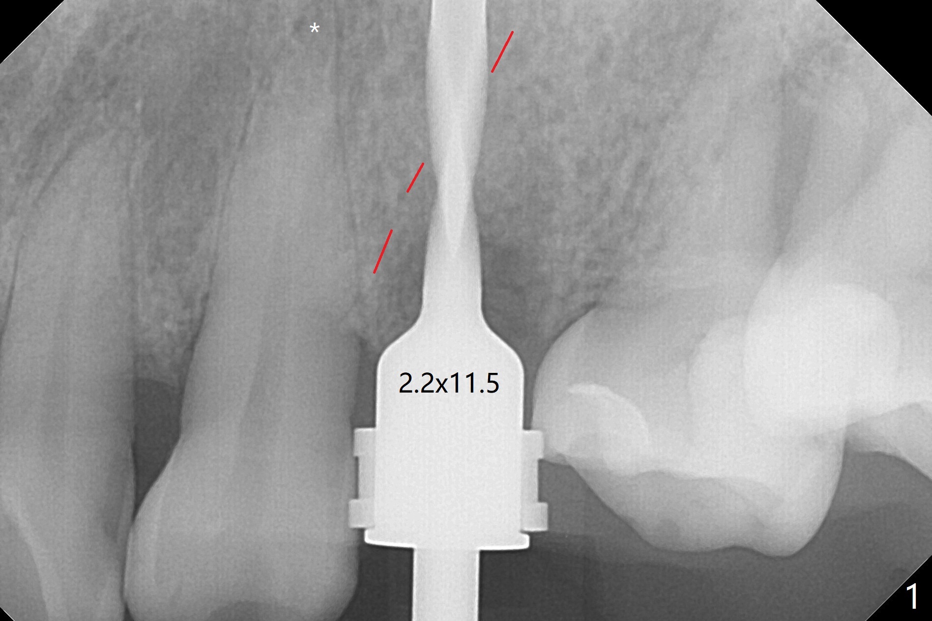
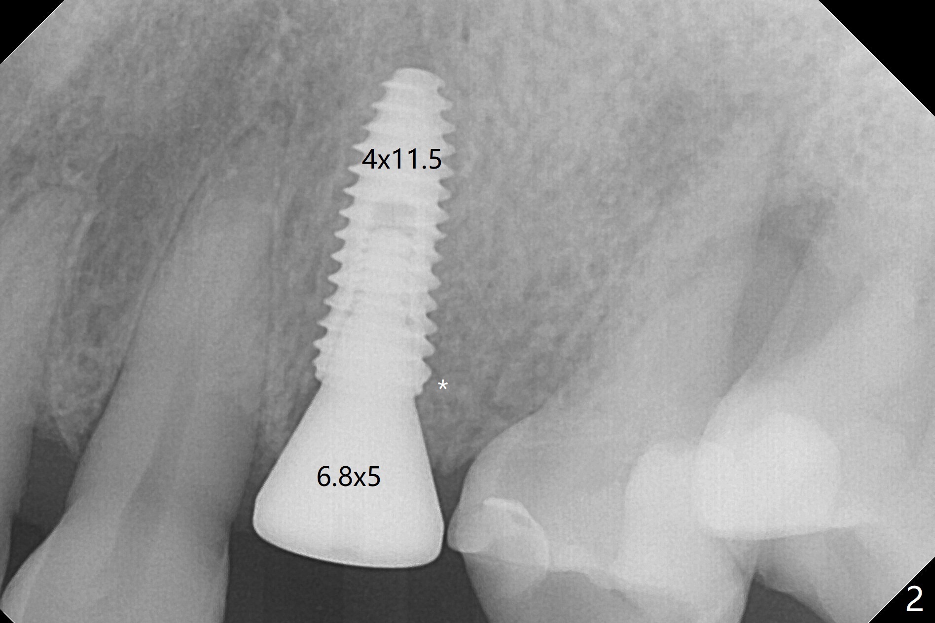
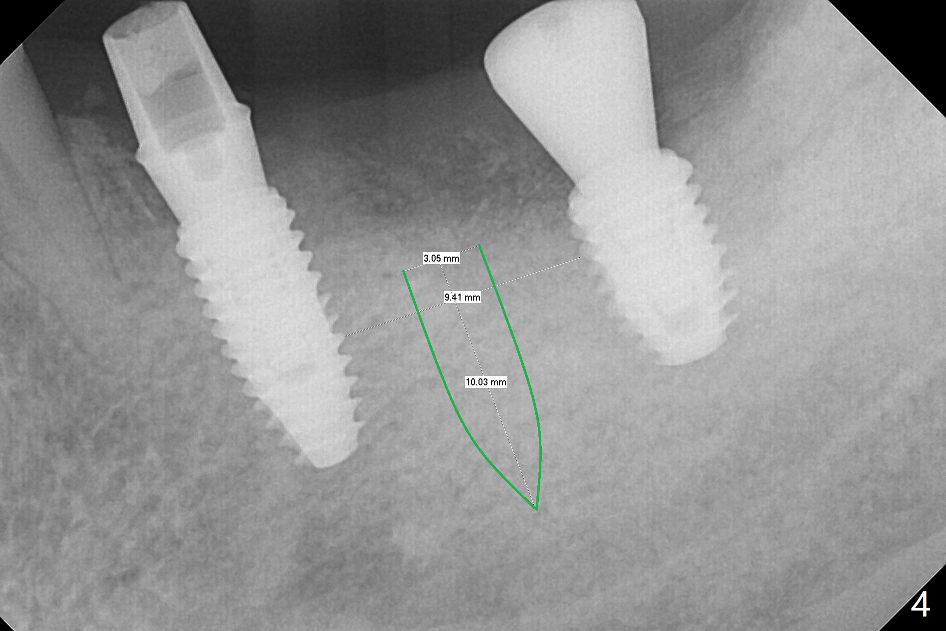
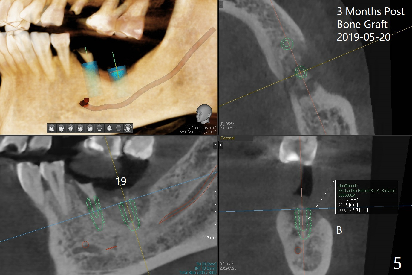
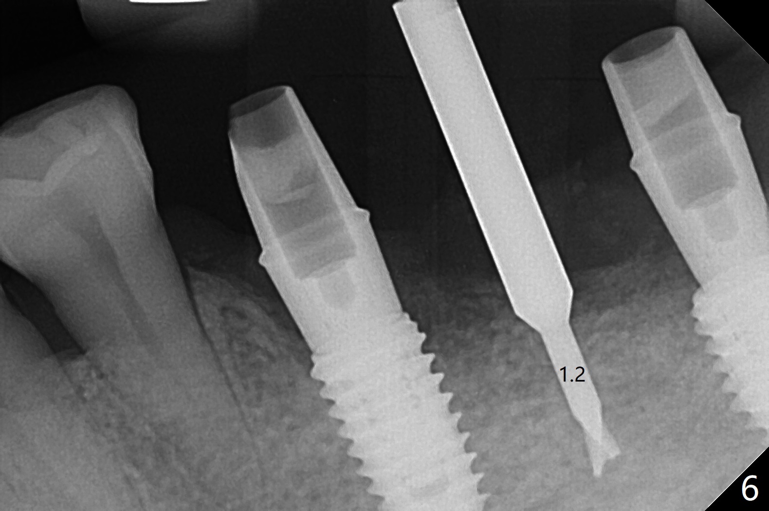
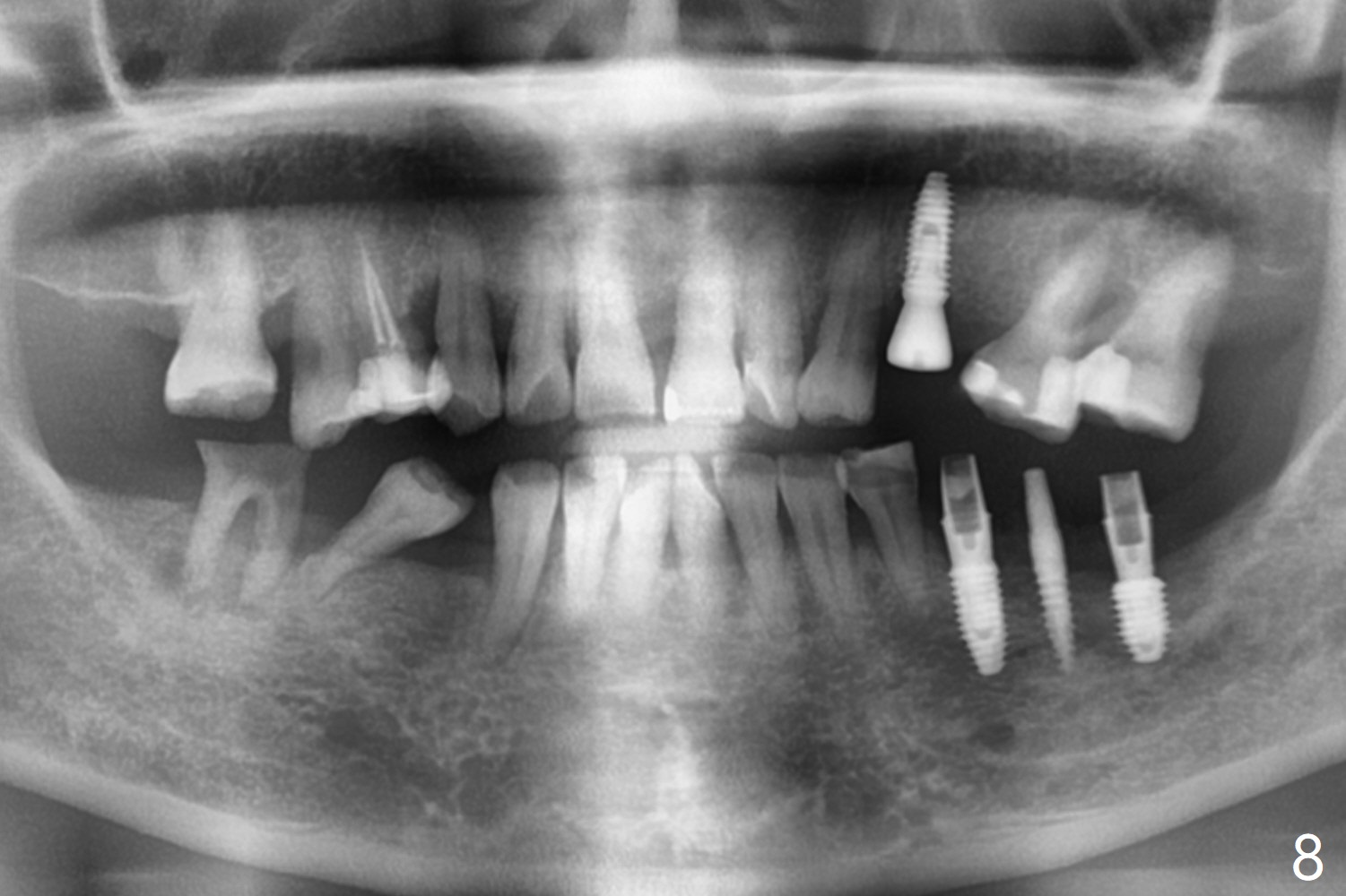
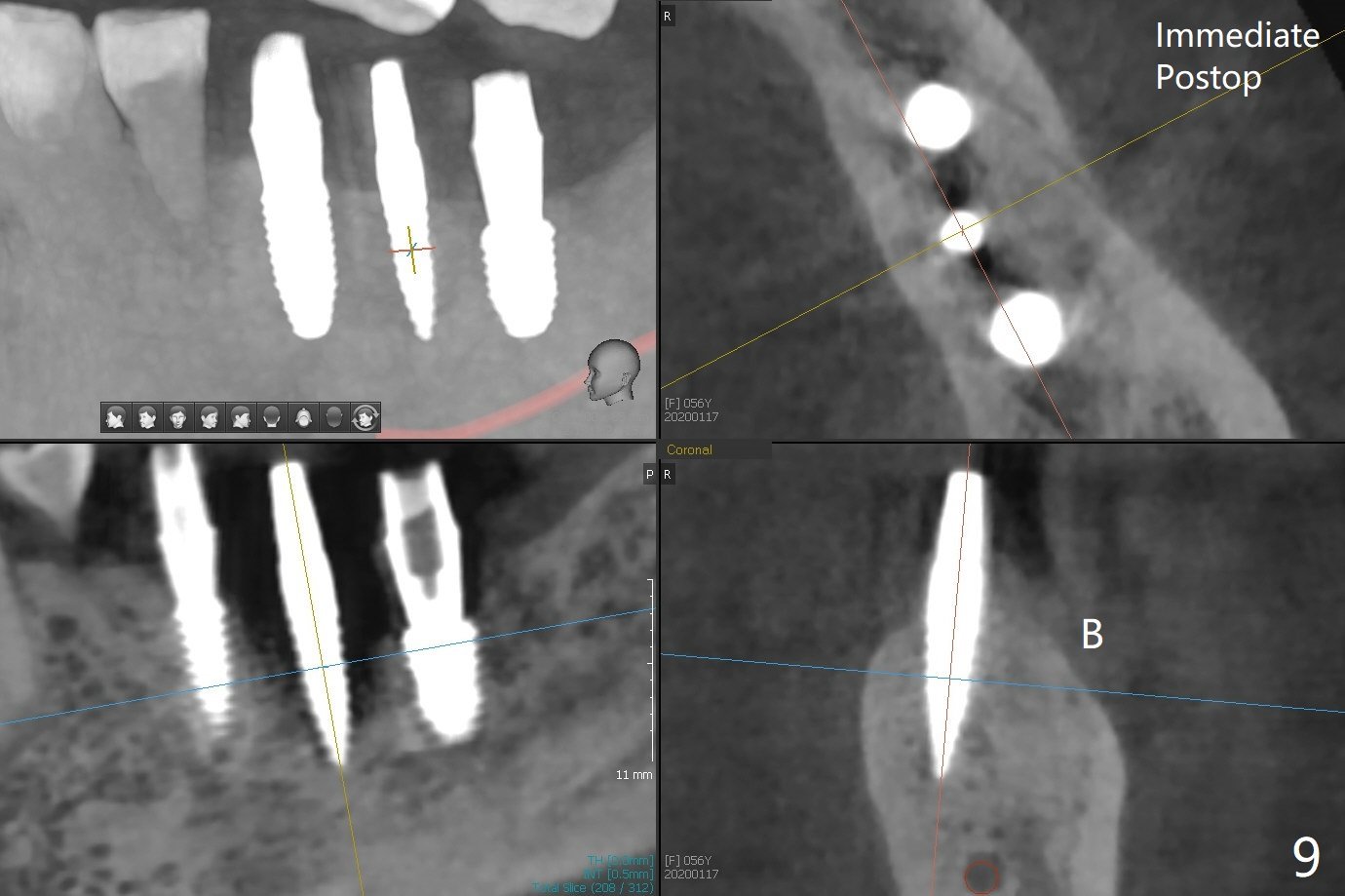
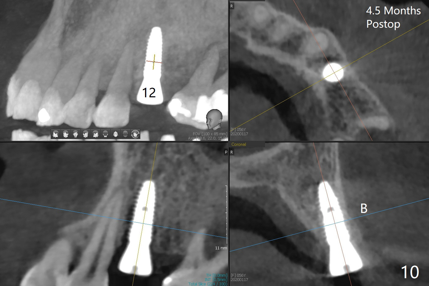
PA is taken after a 2.2 mm drill reaches the depth at #12, since osteotomy is initiated in the mesial slope of the socket (Fig.1 red dashed line) and the neighboring root (*) slightly curves distal. A 8.6x5 mm healing abutment is used to close the socket with the large mesiodistal space after bone graft (Fig.2 *). In contrast, the socket at #20 is large; a 4.5x4.5(3) mm cementation abutment is placed for an immediate provisional to keep autogenous bone (harvested from the site of #18) in place (Fig.3). Six months postop, the patient does not want implant FPD. She wants an additional implant at #19. Since the space between the implants #18 and 20 is 9.41 mm, a narrow implant is indicated (3 or 3.5 mm, Fig.4), in spite of the sufficient buccolingual width (Fig.5). The position and trajectory of the 1.2 mm initial drill and 3x10(2) mm 1-piece implant are acceptable with free hand (Fig.6,7). After 3-4 more turns, panoramic X-ray (Fig.8) and CT (Fig.9) are taken for 28-30 guide. It appears that the 1-piece implant is placed acceptable buccolingual (Fig.9 B). The implants at #12,18,20 (4.5 months postop with guide) are shown in Fig.10-12. It appears that guided surgery is superior in buccolingual position and trajectory to free hand.
Return to
Lower
Molar Immediate Implant,
Trajectory
No Deviation
28-30
Xin Wei, DDS, PhD, MS 1st edition
07/02/2019, last revision
03/21/2020