,%205x7.3,%206x5.jpg)
%20mm,%20later%203-4%20more%20turn.JPG)
.jpg)
|
|
|
|
|
|
|
|
|
|
|
|
|
|
|
|
|
|
Healing Abutment for Small Socket M
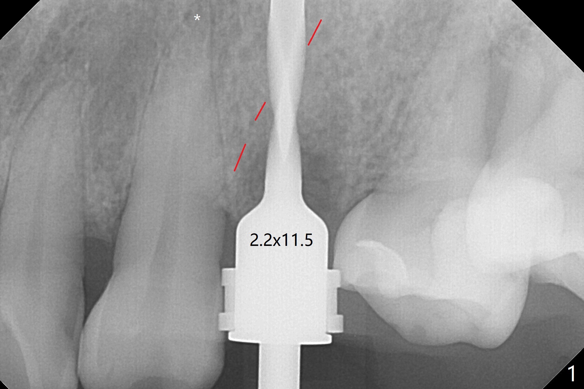
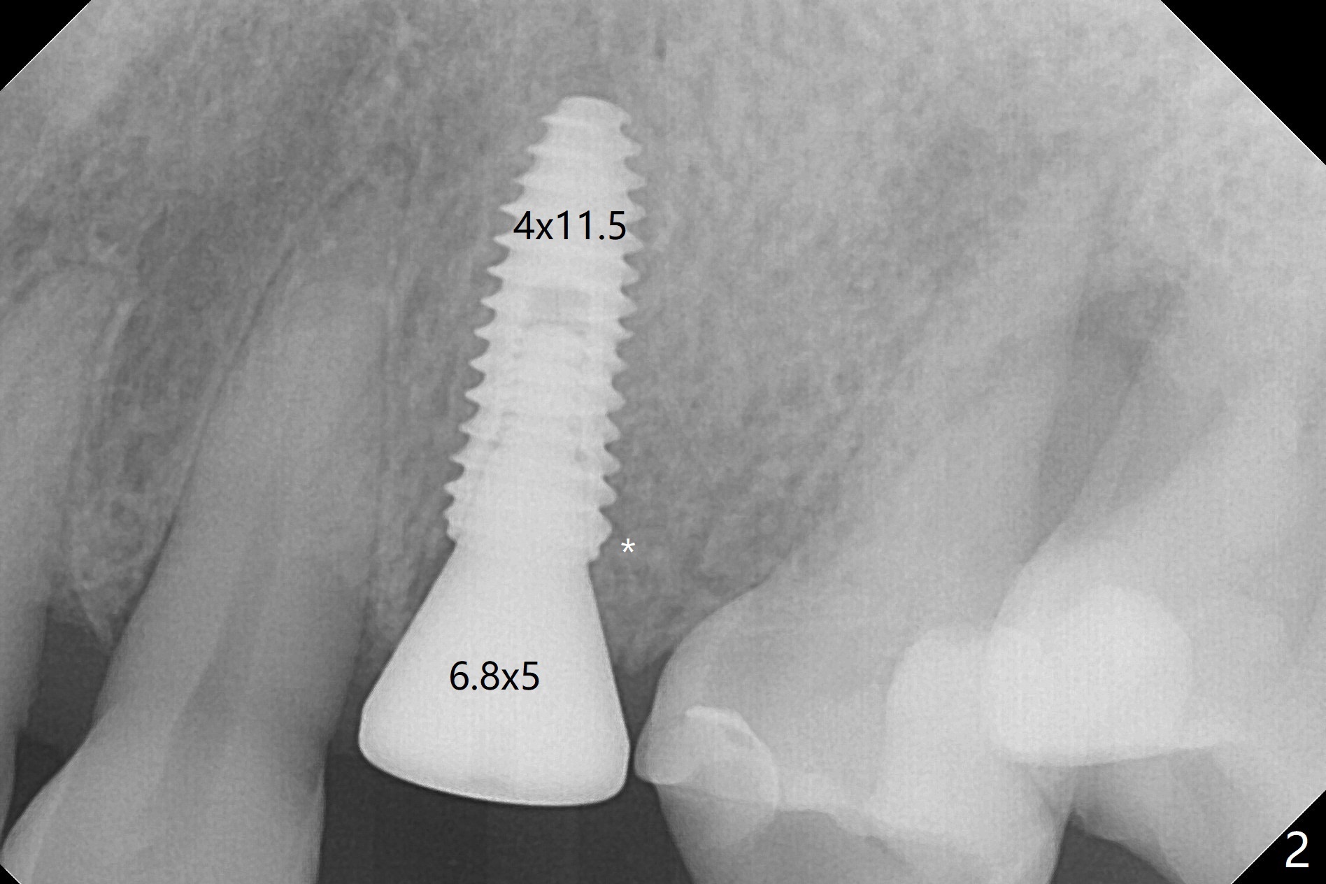
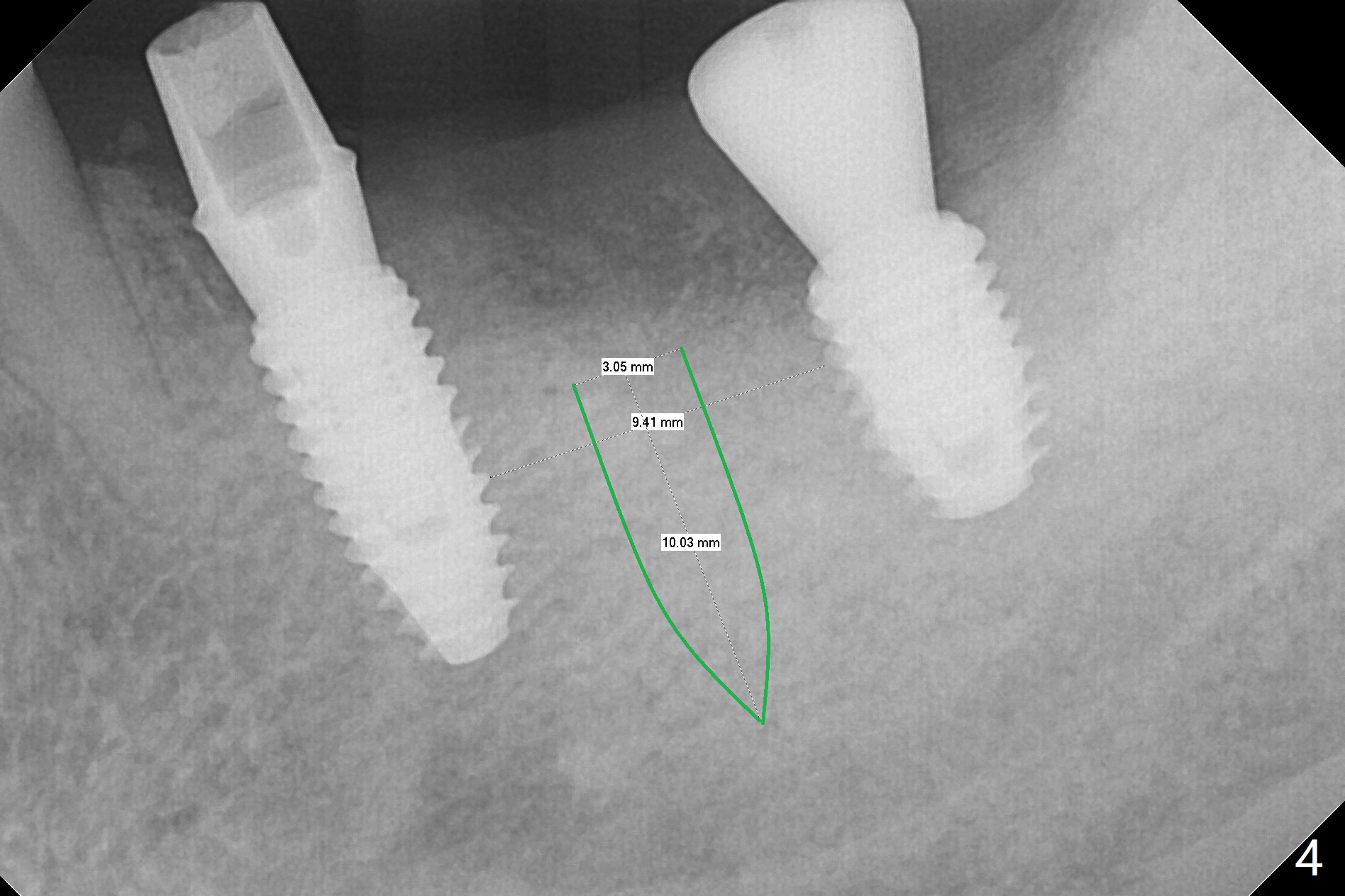
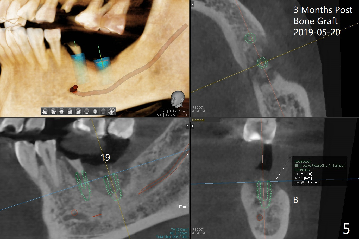
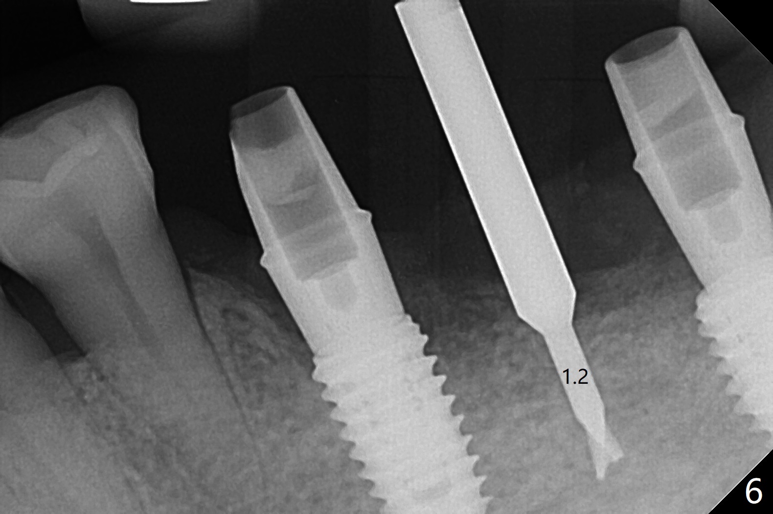
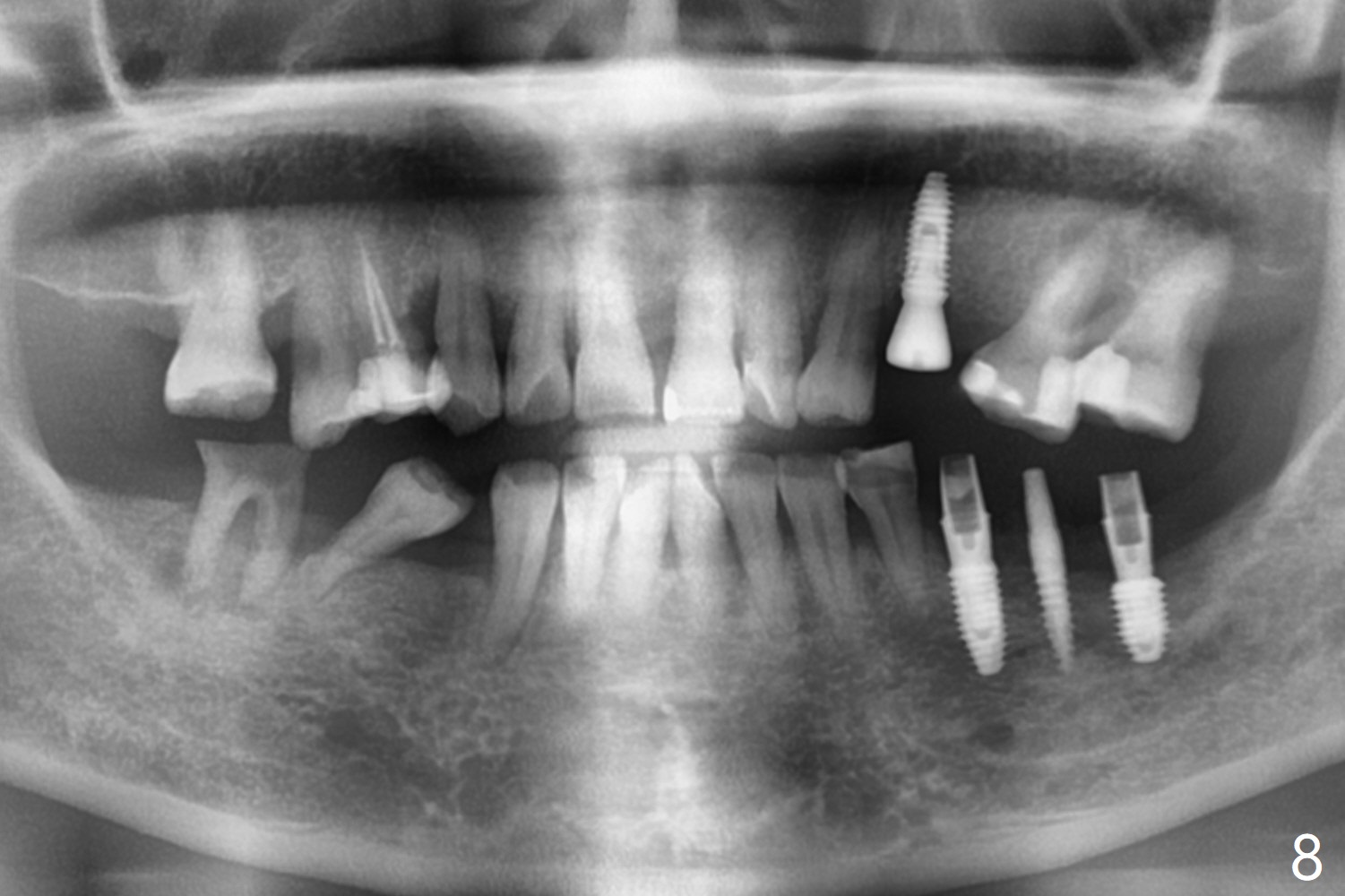
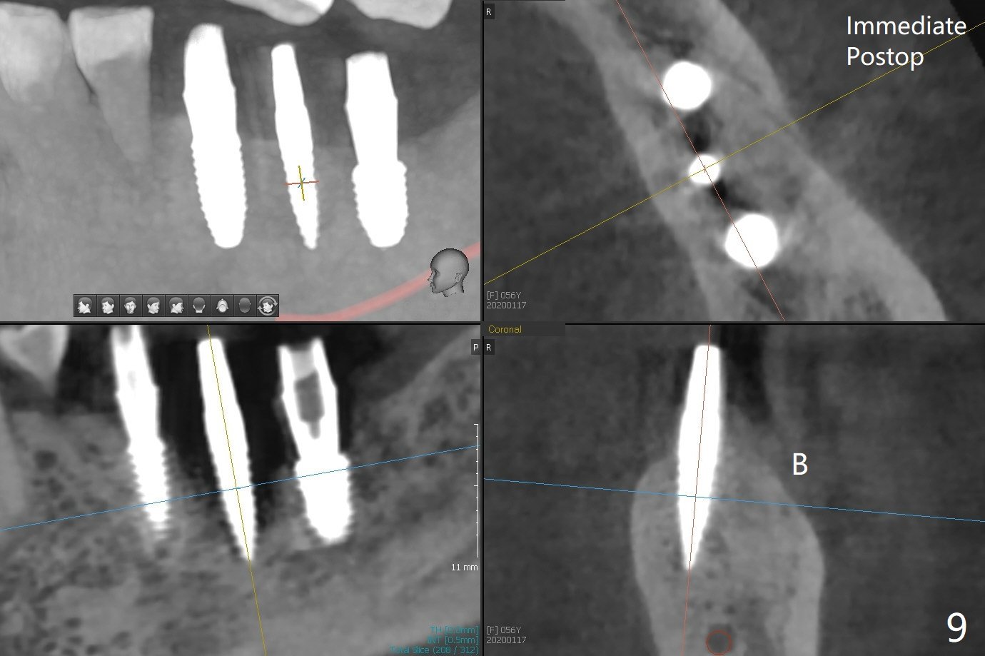
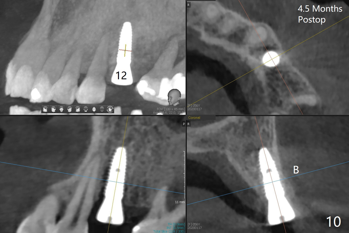
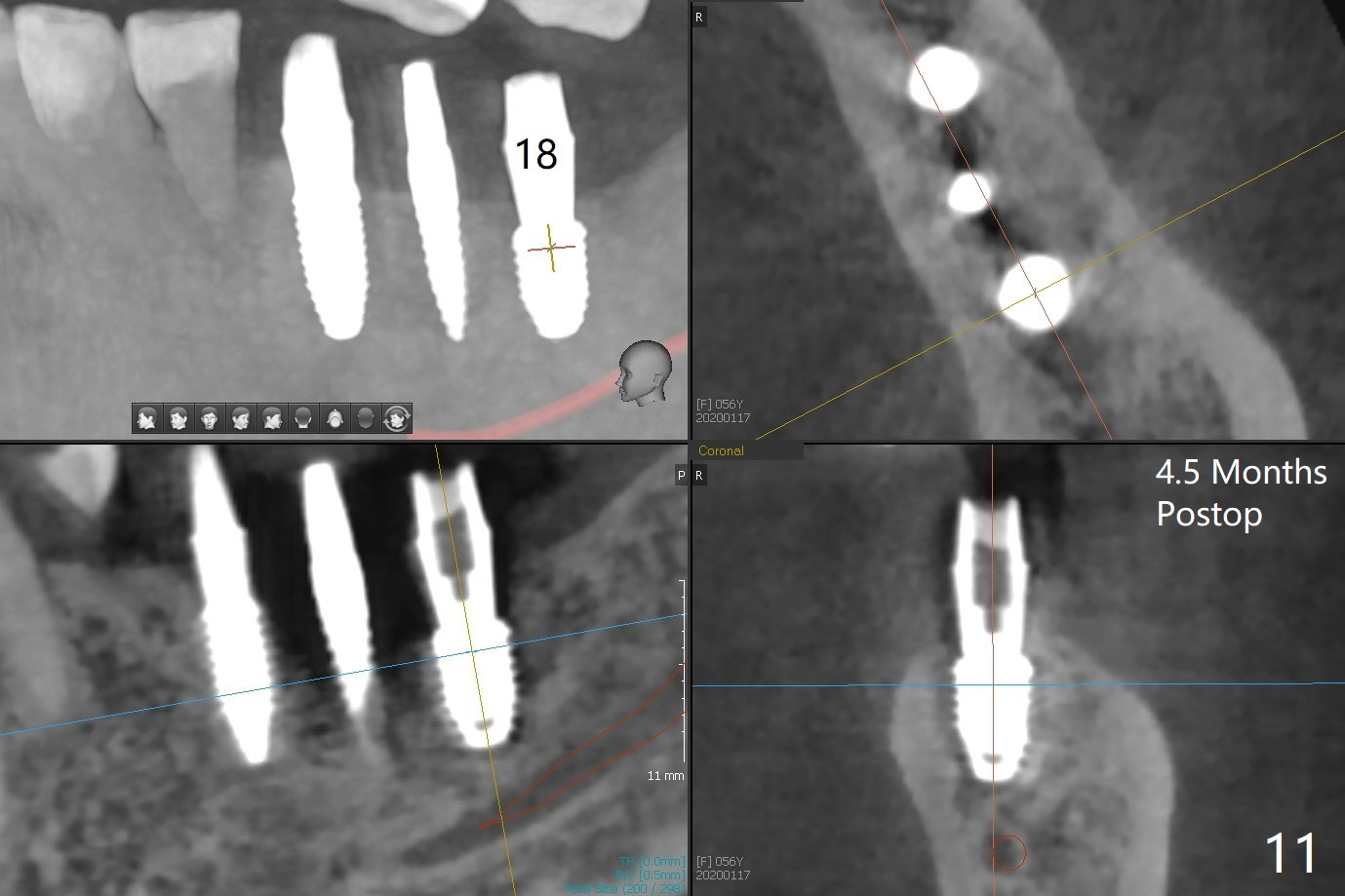
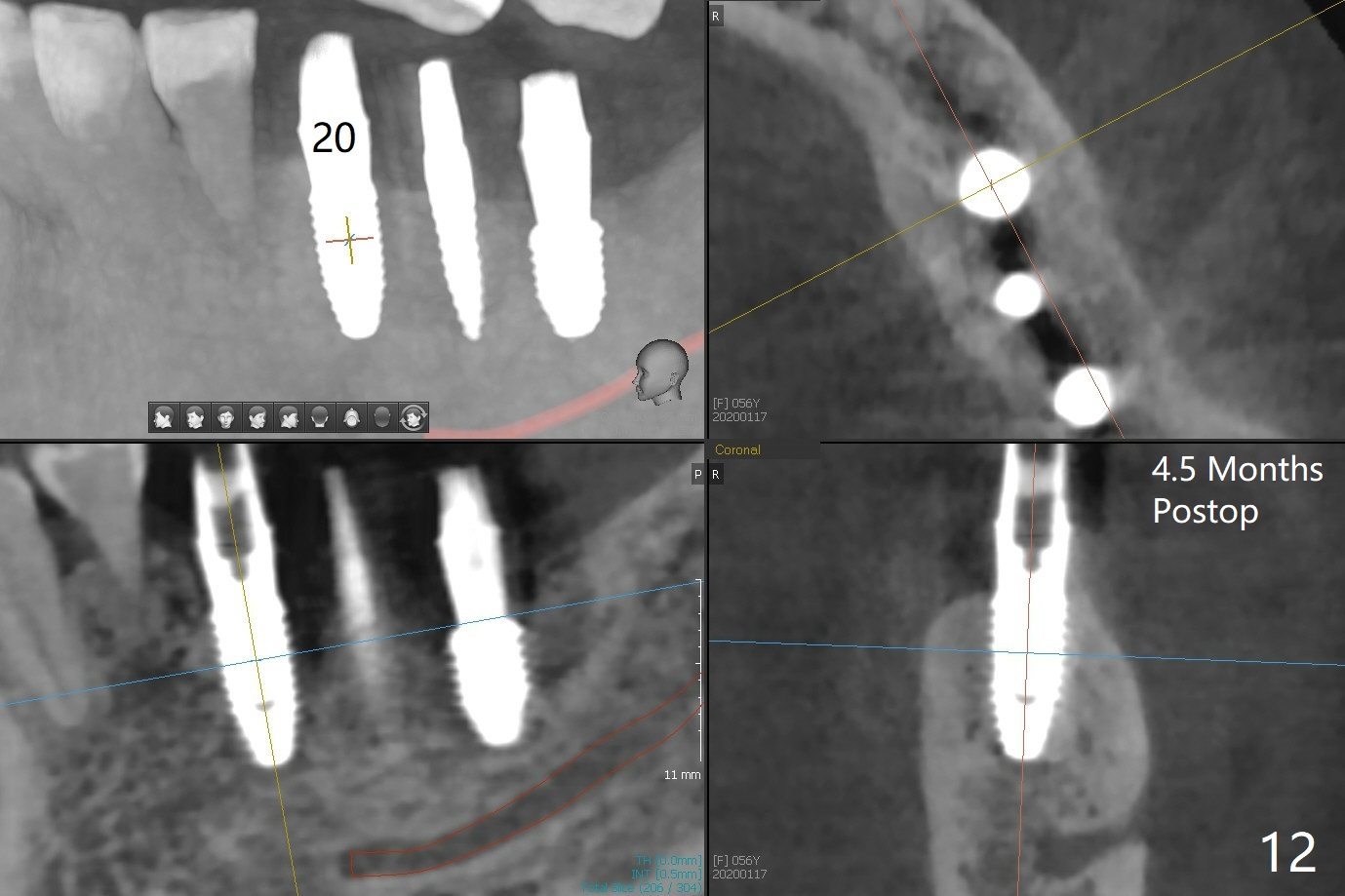
PA is taken after a 2.2 mm drill reaches the depth at #12, since osteotomy is initiated in the mesial slope of the socket (Fig.1 red dashed line) and the neighboring root (*) slightly curves distal. A 8.6x5 mm healing abutment is used to close the socket with the large mesiodistal space after bone graft (Fig.2 *). In contrast, the socket at #20 is large; a 4.5x4.5(3) mm cementation abutment is placed for an immediate provisional to keep autogenous bone (harvested from the site of #18) in place (Fig.3). Six months postop, the patient does not want implant FPD. She wants an additional implant at #19. Since the space between the implants #18 and 20 is 9.41 mm, a narrow implant is indicated (3 or 3.5 mm, Fig.4), in spite of the sufficient buccolingual width (Fig.5). The position and trajectory of the 1.2 mm initial drill and 3x10(2) mm 1-piece implant are acceptable with free hand (Fig.6,7). After 3-4 more turns, panoramic X-ray (Fig.8) and CT (Fig.9) are taken for 28-30 guide. It appears that the 1-piece implant is placed acceptable buccolingual (Fig.9 B). The implants at #12,18,20 (4.5 months postop with guide) are shown in Fig.10-12. It appears that guided surgery is superior in buccolingual position and trajectory to free hand. There is crestal remodeling without implant thread exposure 11 months postop (Fig.13).
Return to
Lower
Molar Immediate Implant,
Trajectory
No Deviation
28-30
Xin Wei, DDS, PhD, MS 1st edition
07/02/2019, last revision
06/09/2020