|
|
|
|
|
|
|
|
|
|
|
|
|
||
|
|
|
|
|
|
|
|
||
Redo I
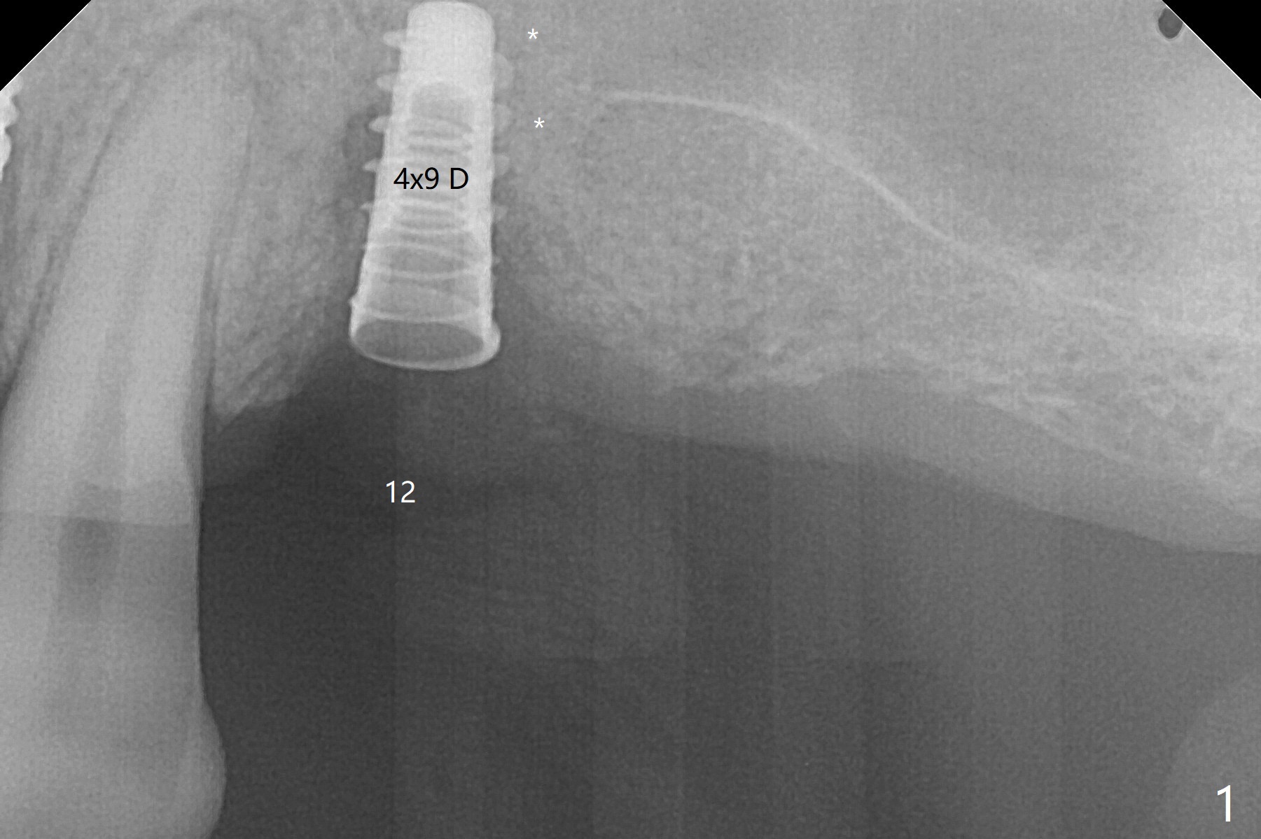
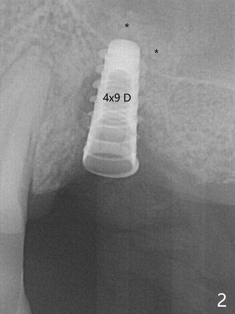
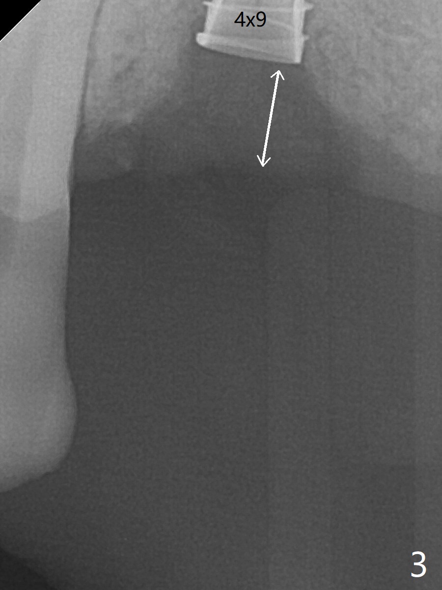
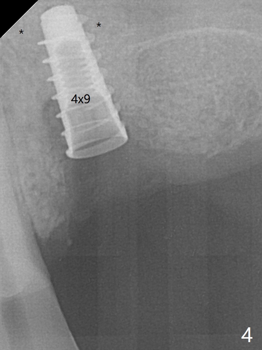
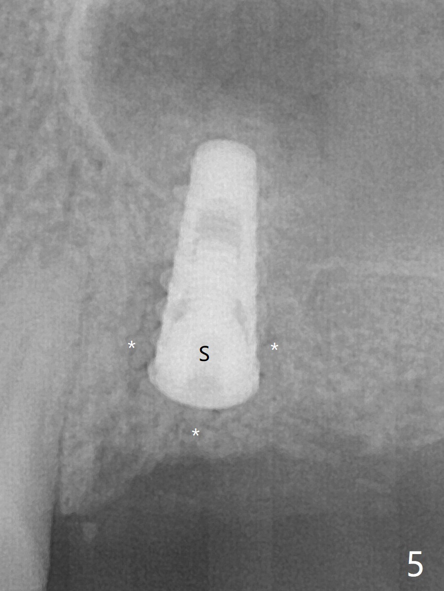
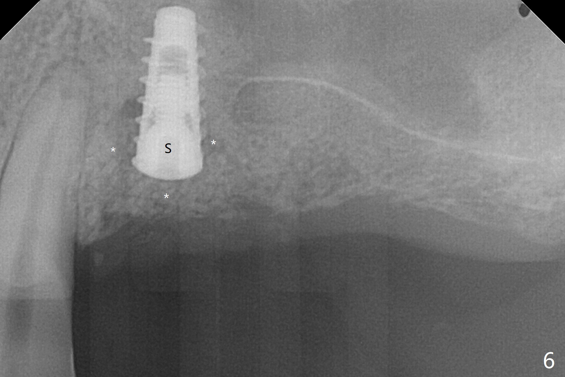
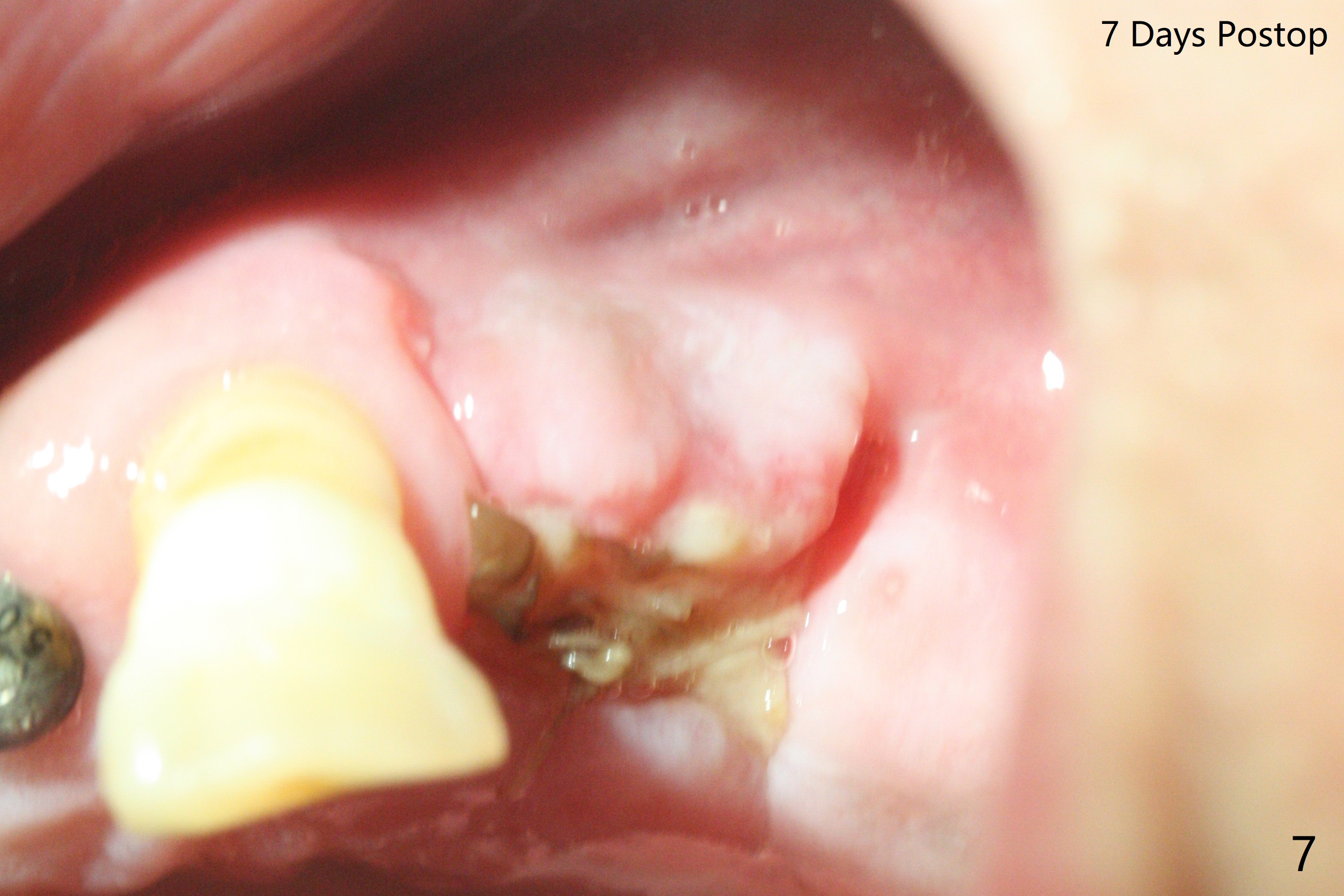
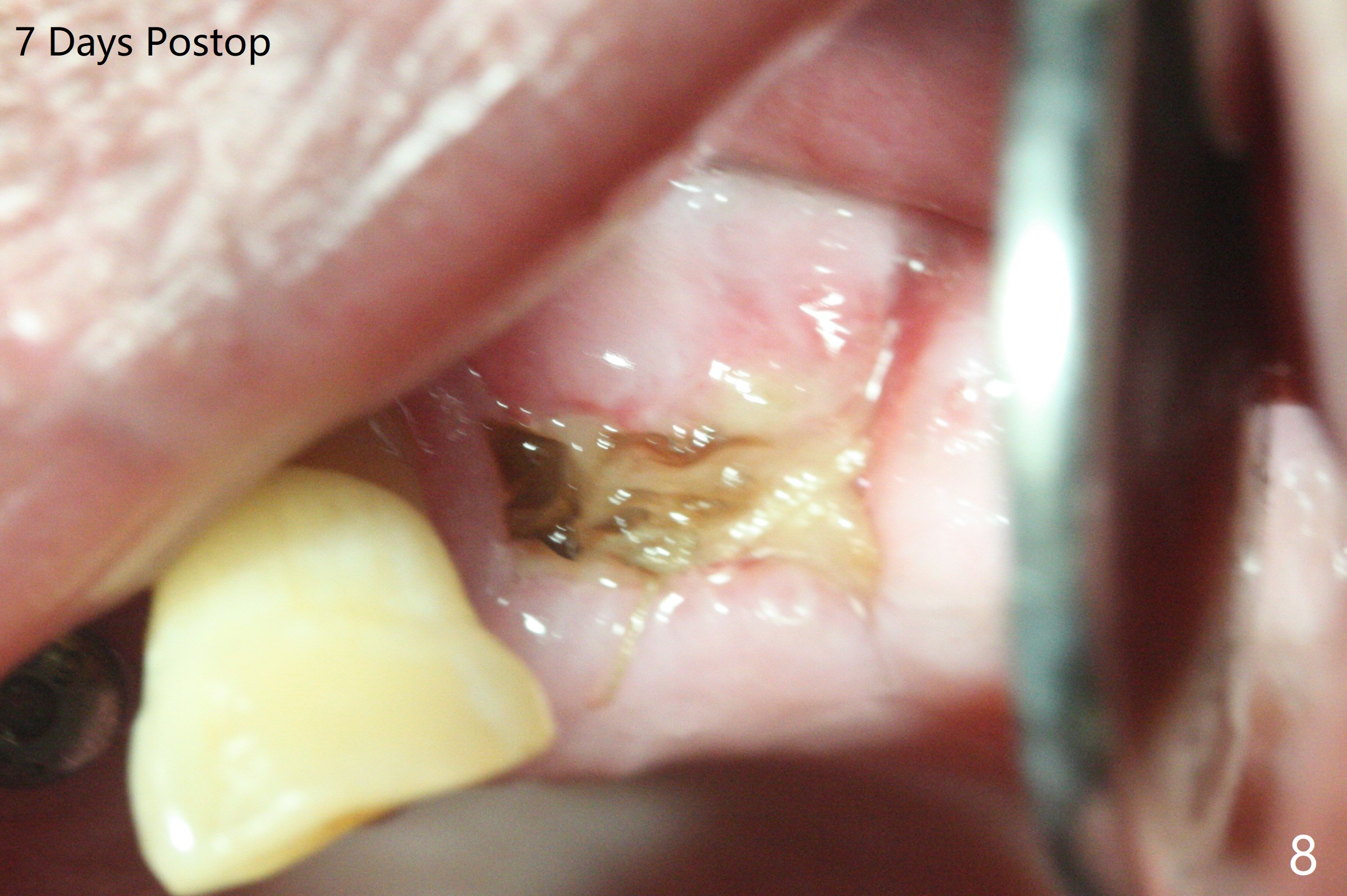
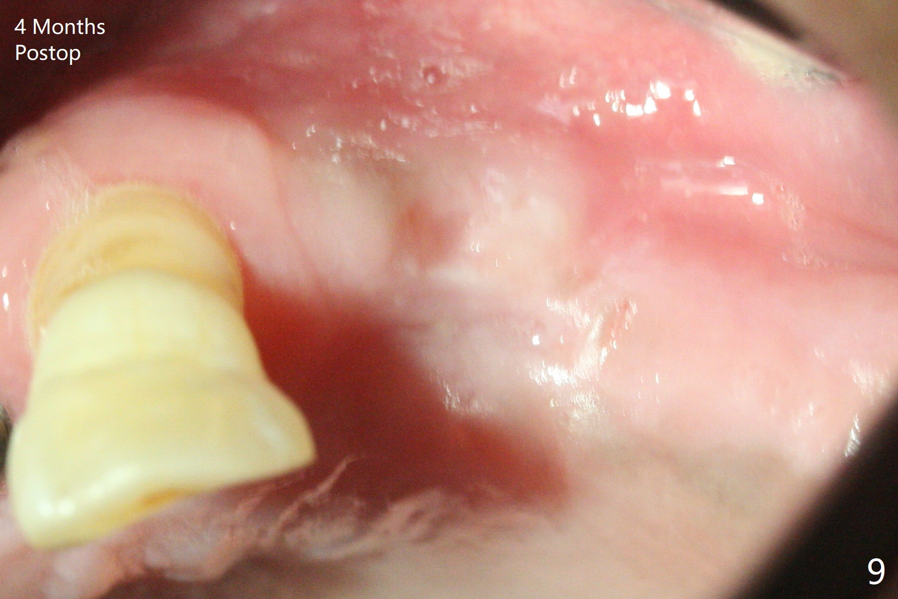
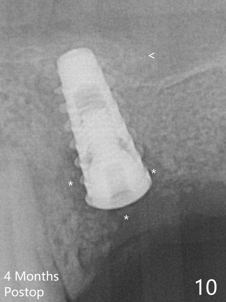
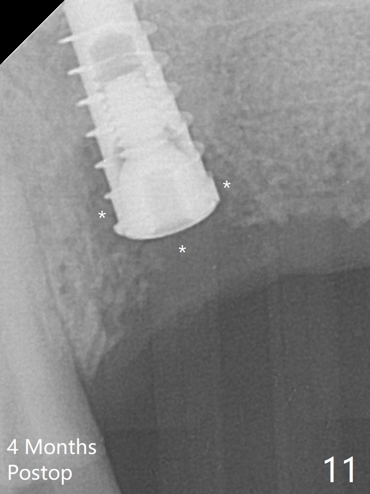
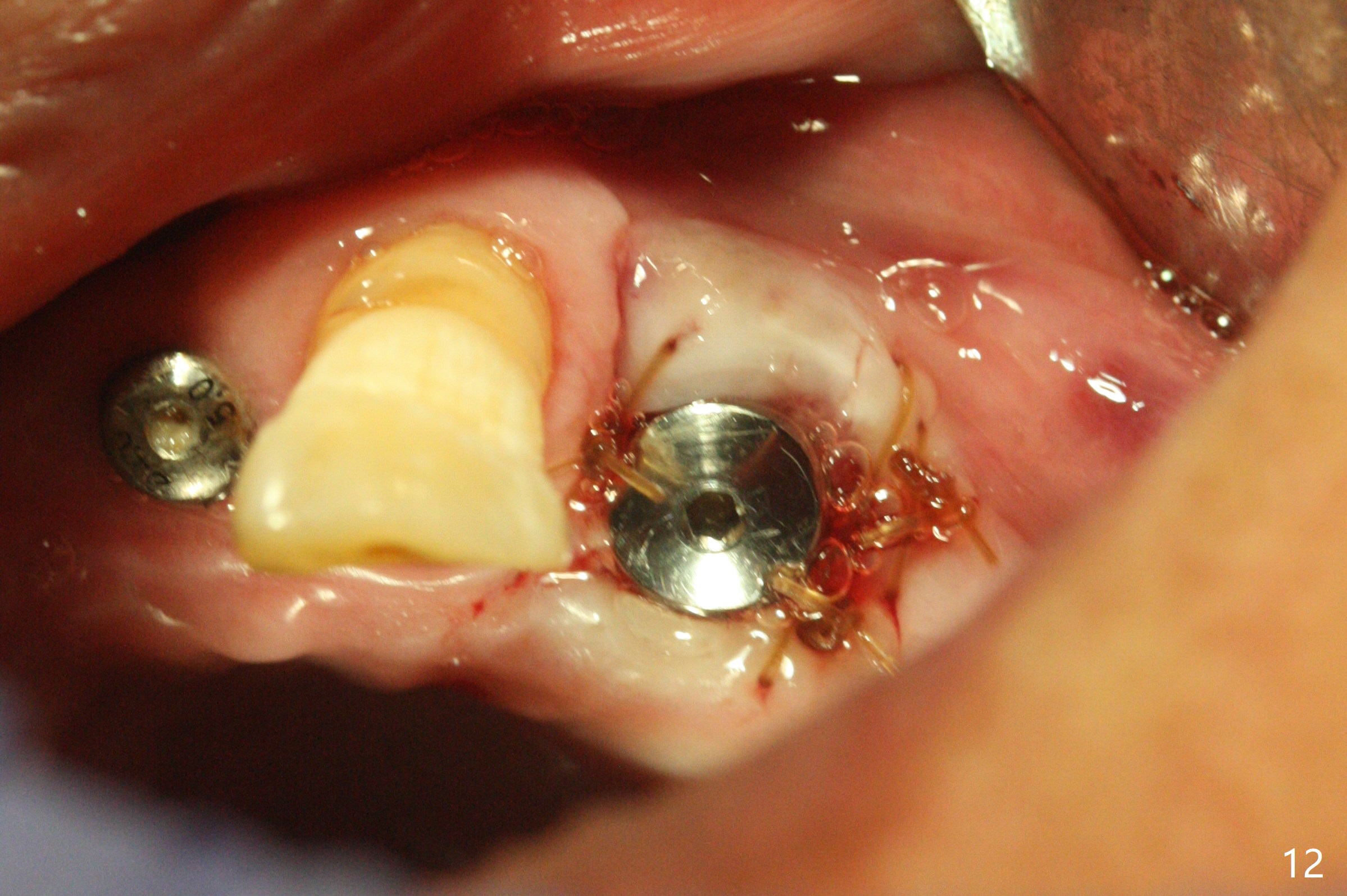
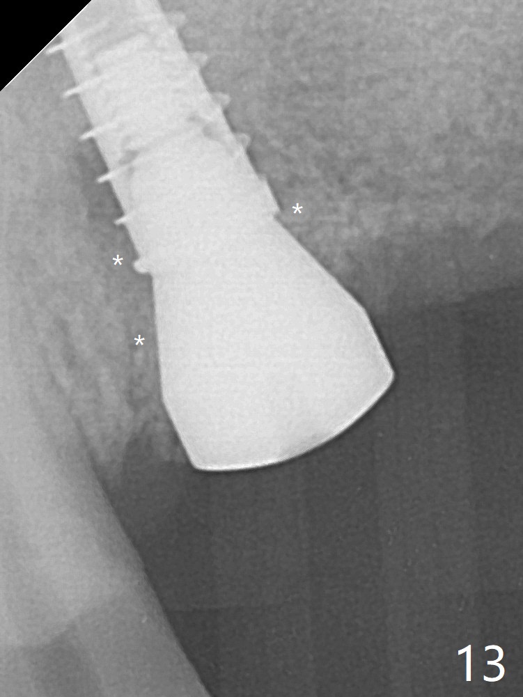
Five months post implant removal and bone graft, incision is made at #12. Osteotomy is being done with guide until 3.0x7.3 mm drill with 1 O-ring (palatal wall being thin), followed by 3.2x17-19 mm sinus round drills and 2.2x11.5 mm drill. After placement of 2 small loads of bone graft, a 4x9 mm dummy implant is inserted for sinus lift (Fig.1,2 *). By this time, the buccal plate is gone, while there is apparently the palatal periosteum. With more bone graft for sinus lift, a 4x9 mm final implant is placed with 30 Ncm (machine) and 4 mm subgingival (Fig.3,4 double arrows). Sticky bone (Fig.5,6 *) is applied around the coronal end of the implant and cover screw (S), followed by 2 pieces of PRF and 4-0 PGA suture. The sutures appear to have been dissolved and PRF membrane exposed 7 days postop (Fig.7,8 (smoker)). Four months postop, the wound heals except a small hole, which seems to be communicated with the underlying implant (Fig.9). The sinus lift remains (Fig.10 <), while bone loss appears to be present around the implant (Fig10,11 *). After placement of 5.5x4 mm healing abutment and before suturing, allograft is pushed into periimplant space (Fig.12, 13 *).
Return to
Trajectory
No Deviation
25
Xin Wei, DDS, PhD, MS 1st edition
02/13/2020, last revision
06/24/2020