,%20native%20bone%204.8%20mm.jpg)
|
|
|
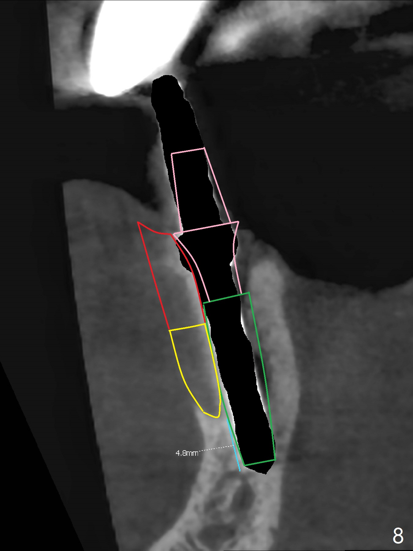
After implant placement (Fig.8 (CT coronal section) green), the apical portion of the buccal gap is filled with Osteogen plug (yellow). Post abutment insertion (pink), allograft (red in Fig.8; * in Fig.9) is placed around the coronal implant and the cuff of the abutment.
2-Pointed Fixation Last Next Xin Wei, DDS, PhD, MS 1st edition 01/10/2019, last revision 01/11/2019