,%20Vanilla.jpg)

|
|
|
|
|
|
|
|
|
|
|
|
|
|
|
Free Hand Placement
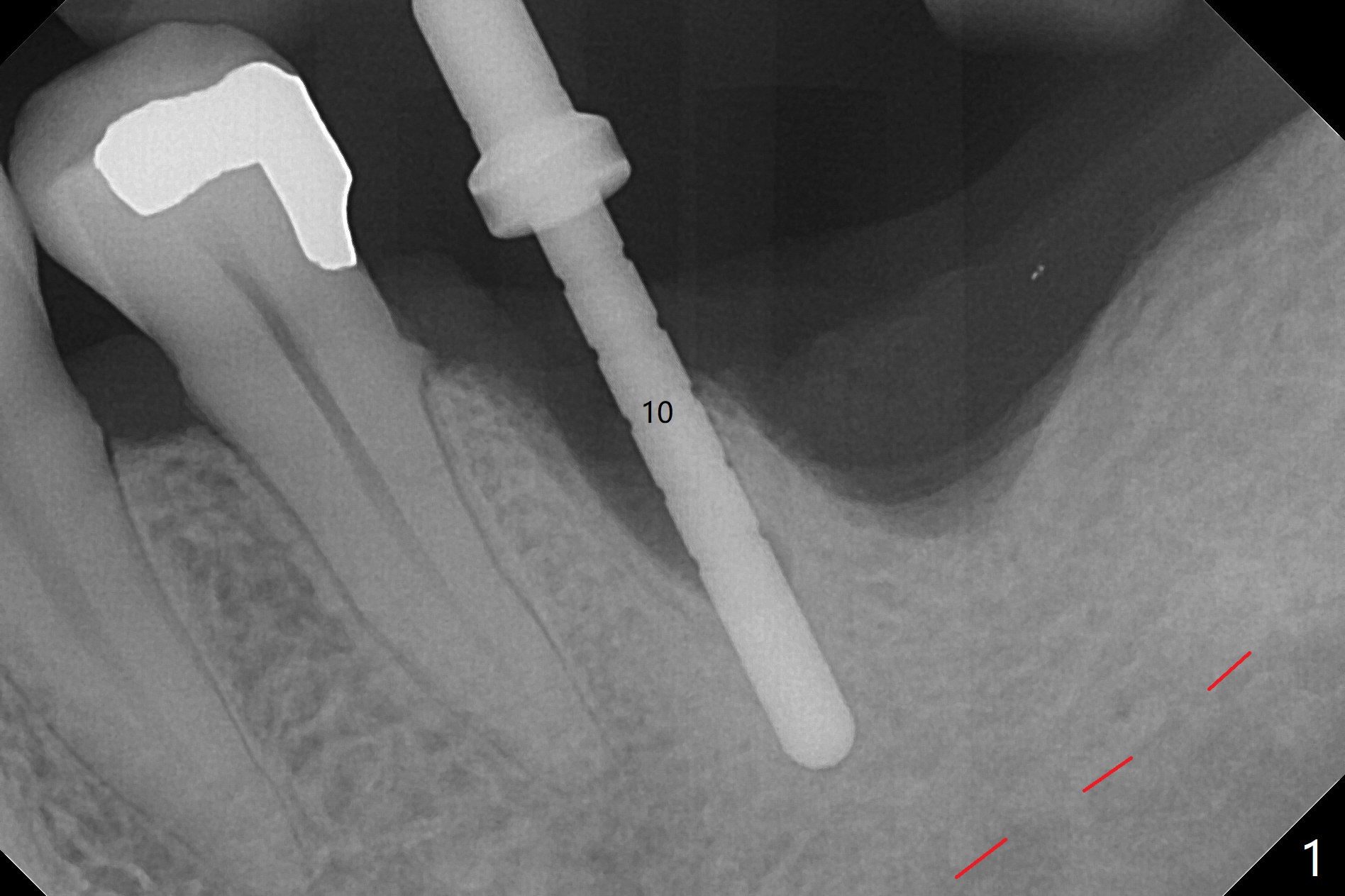
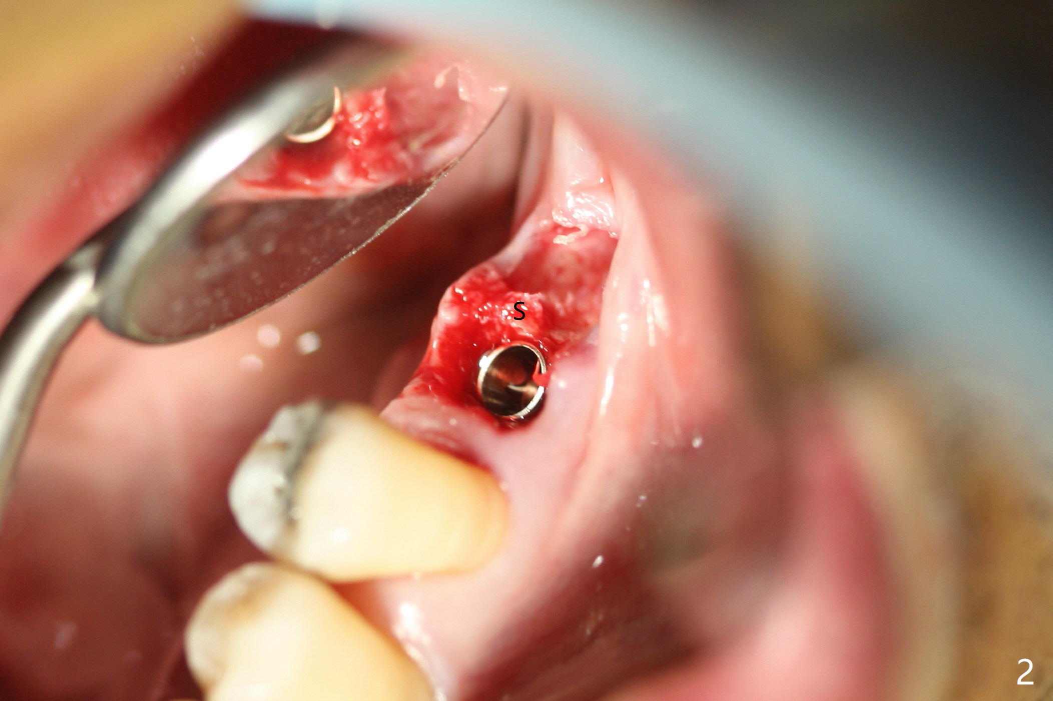
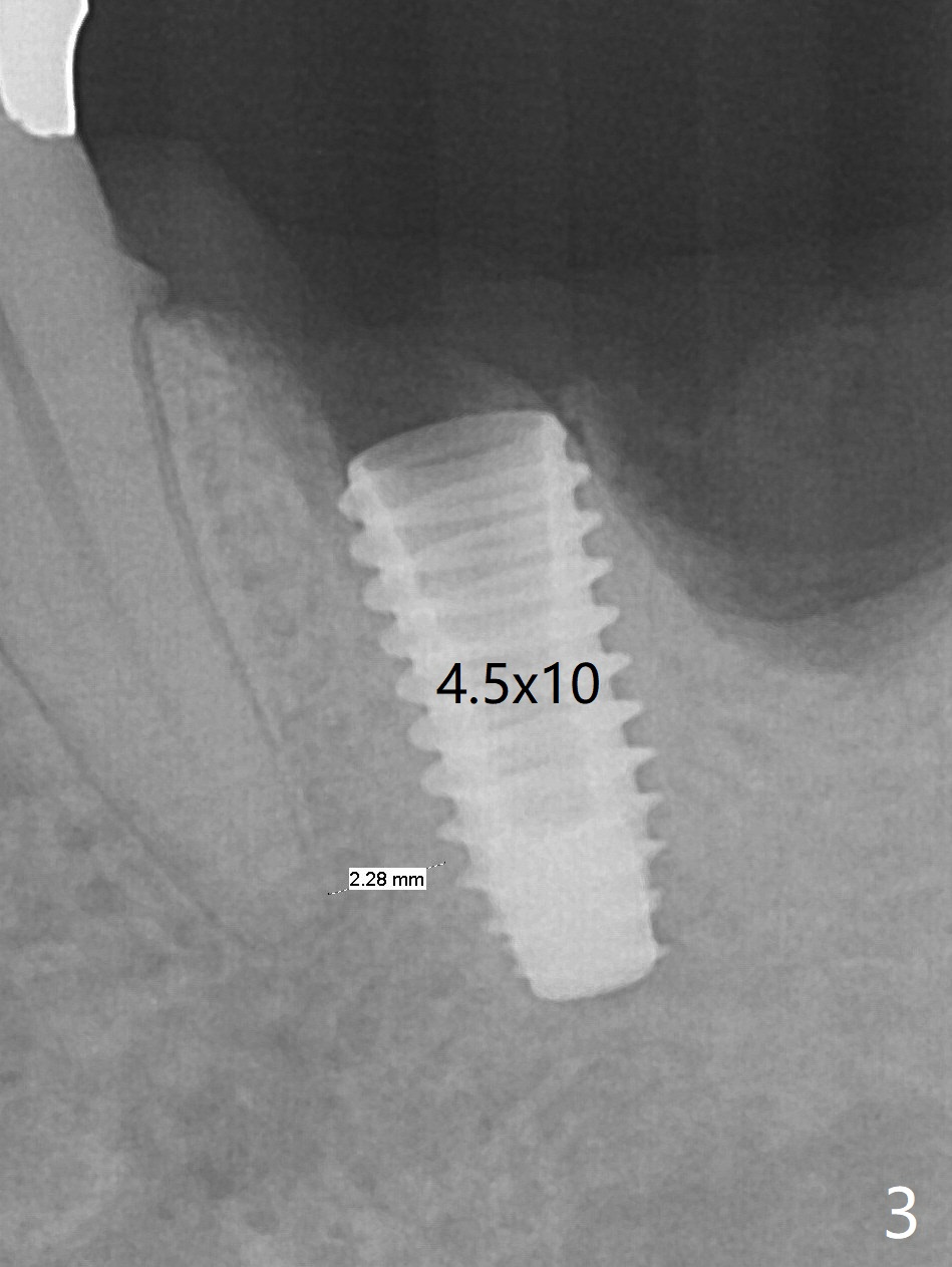
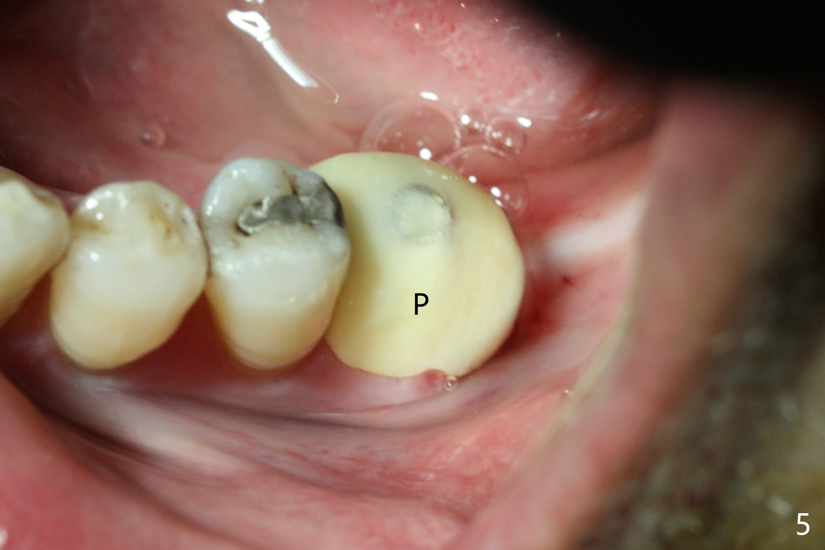
The guide does not arrive when the tooth #19 is extracted. Osteotomy is initiated free hand in the mesial socket as planned, slightly lingual, for 11.5 mm (Fig.1). Following sequential osteotomy, a 4.5x10 mm implant is placed subcrestal septally (Fig.2 S) and 2.3 mm from the neighboring apex (Fig.4). A 5.5x5(4) mm abutment is placed immediately and allograft is placed in the remaining sockets (Fig.4 *). An immediate provisional is fabricated to keep the graft in place (Fig.5 P; the most secure socket preservation).
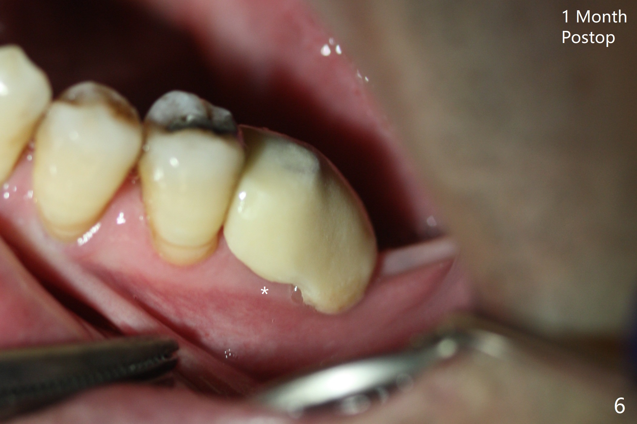
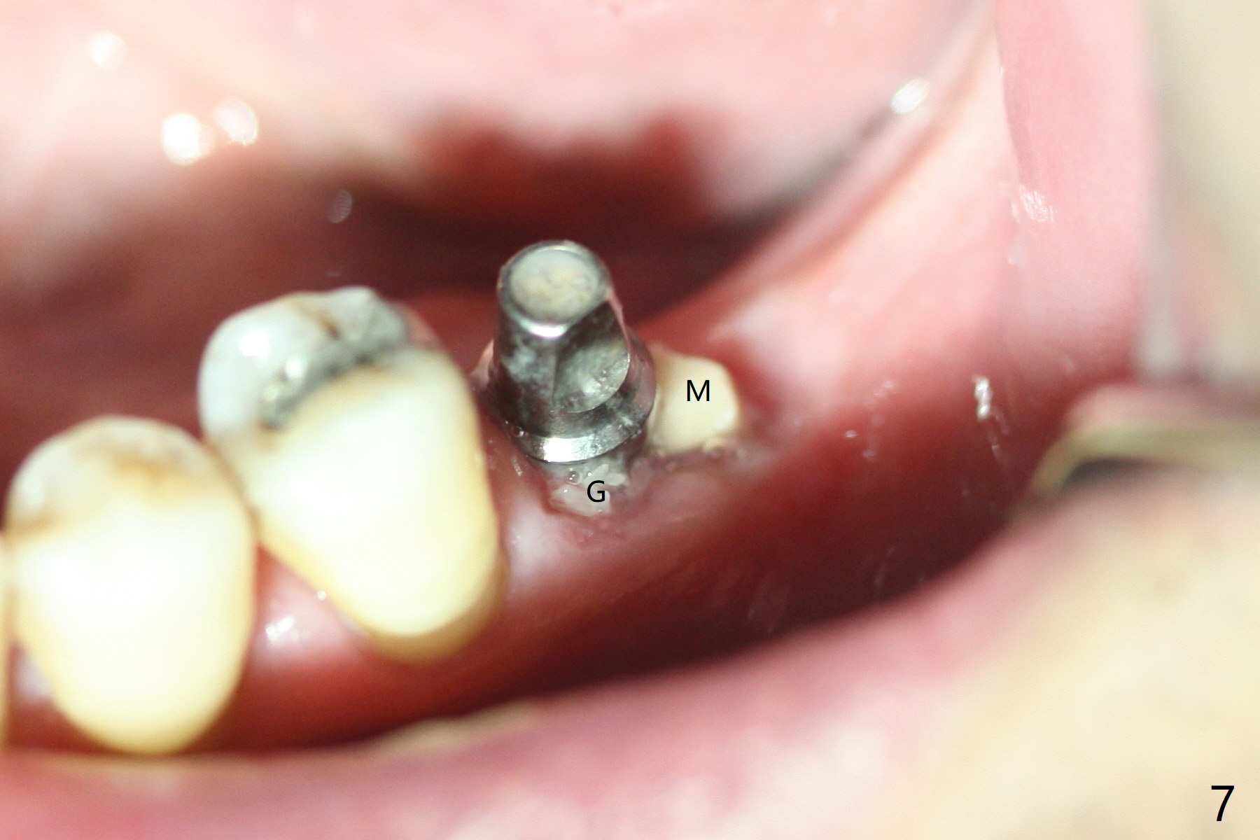
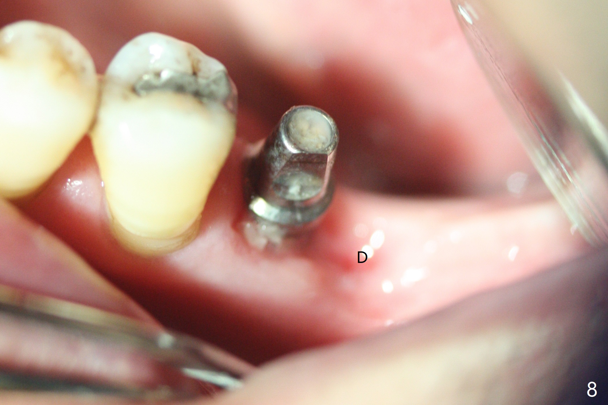
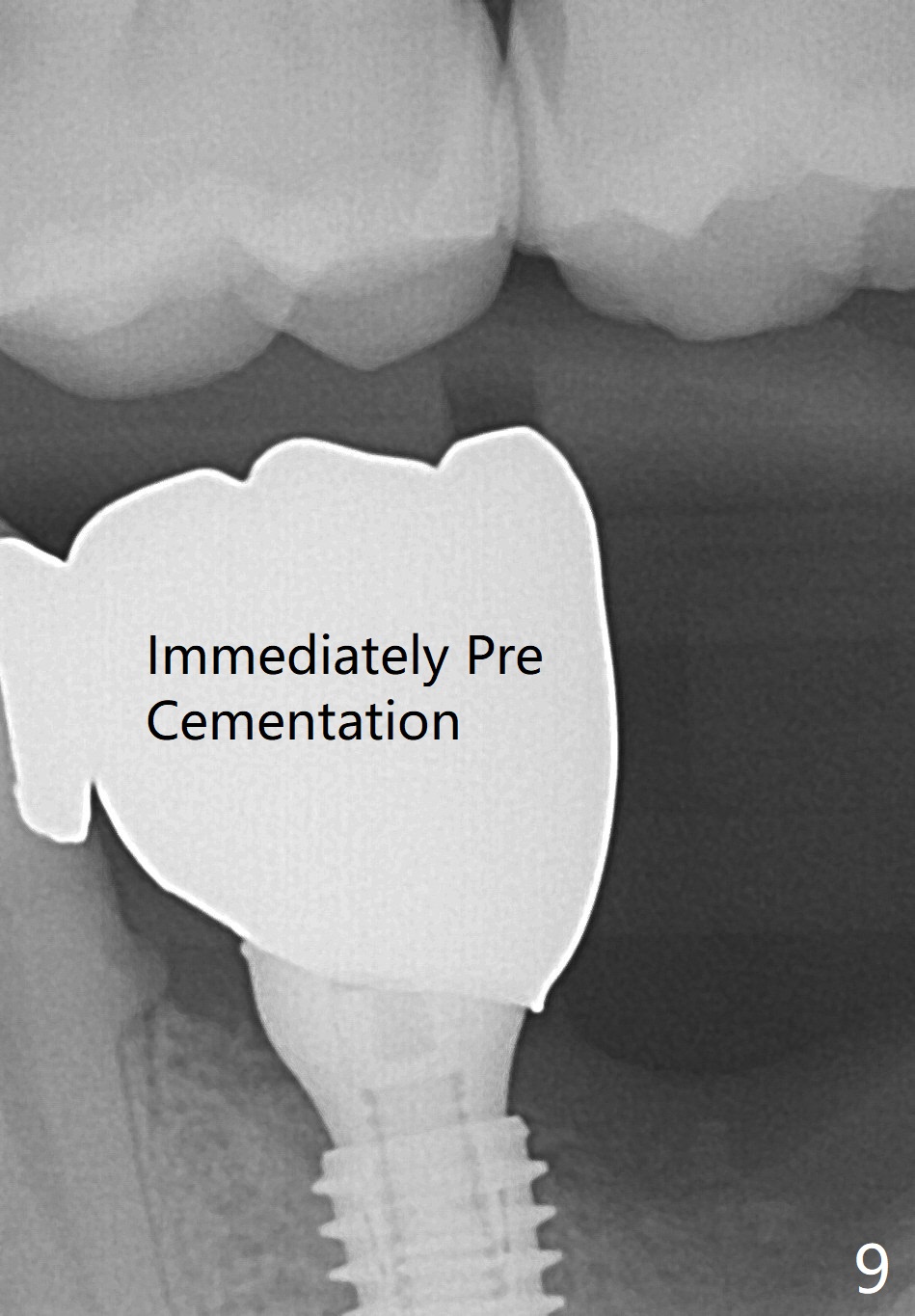
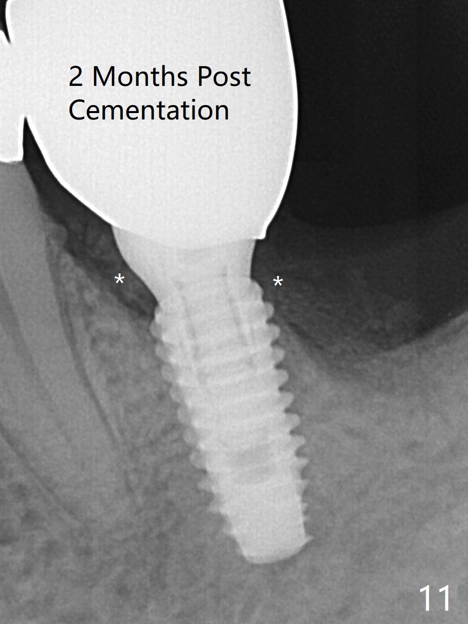
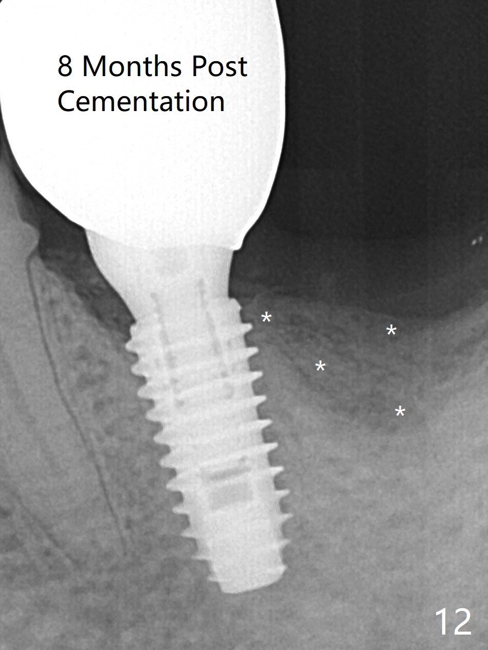
There is shrinkage in the mesial alveolus 1 month postop (Fig.6 *). When the provisional is removed 2 months postop, there is minimal amount of unincorporated bone graft (Fig.7 G) and Osteogen Plug (M, membrane). As the un-attached materials are washed up, the distal socket heals without exposure of implant threads (Fig.8 D). The bone graft settles down and remains in the distal socket 2.5 months postop (immediately pre-cementation, Fig.10 *). The bone graft appears to prevent implant threads from being exposed 2 months post cementation (Fig.11). The bone density in the distal socket increases significantly 8 months post cementation (Fig.12 *).
Return to
Lower
Molar Immediate Implant,
Prevent Molar Periimplantitis (Protocols,
Table),
Trajectory
Xin Wei, DDS, PhD, MS 1st edition 12/13/2018, last revision 11/09/2019