|
|
|
|
|
|
|
|
|
|
Bone or Tissue-Level Implant?
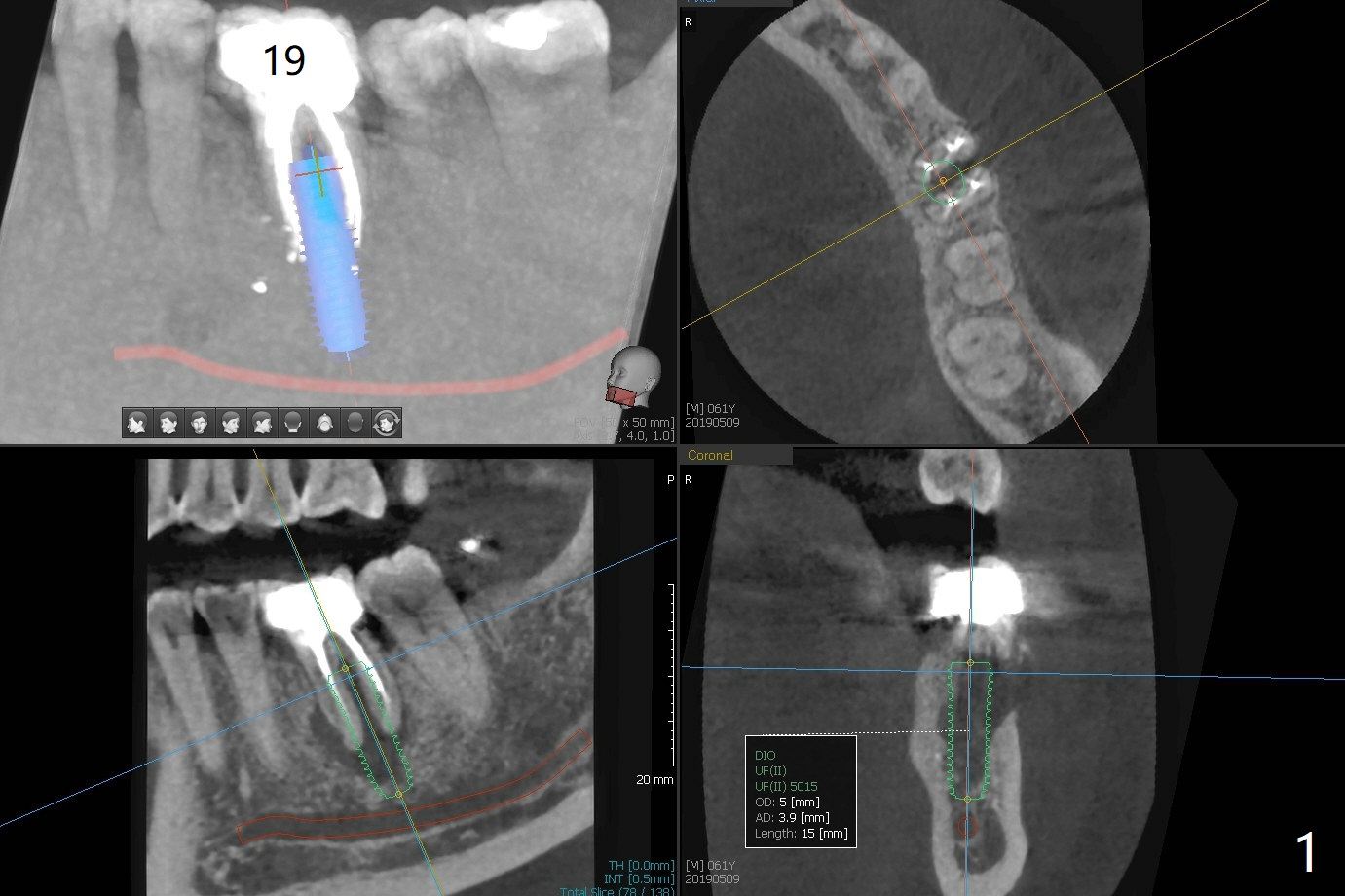
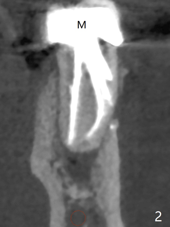
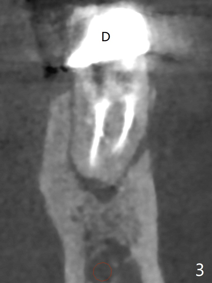
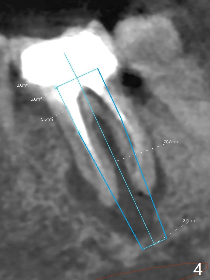
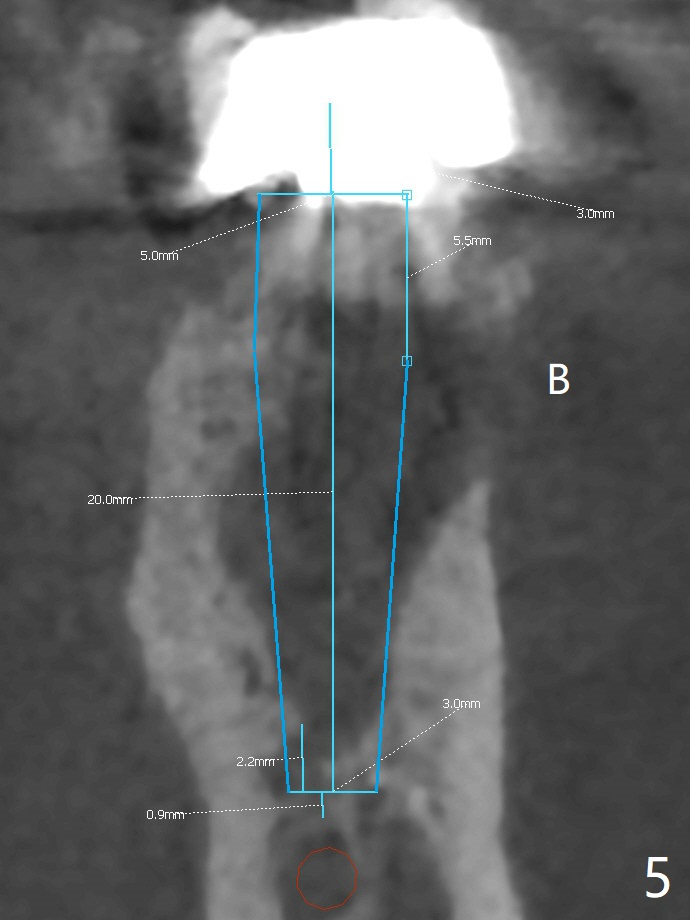
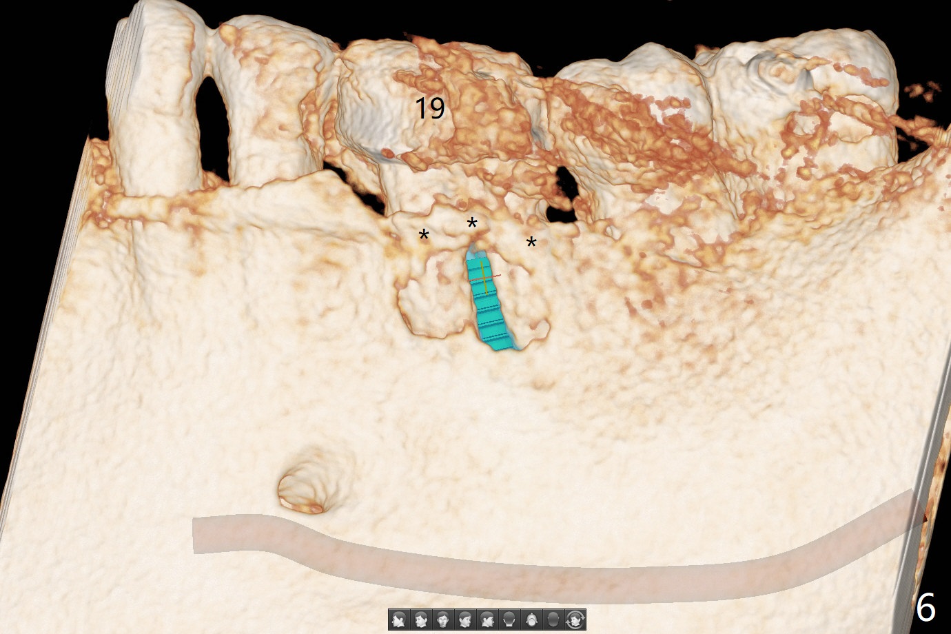
The tooth #19 of a 61-year-old man develops pain 9 years post complicated RCT (Fig.1). Periradicular radiolucency is more around the mesial root (Fig.2) than the distal one (Fig.3). Section the crown to check whether the distal margin is restorable. Remove the mesial root to determine whether the distal one is salvageable or not. If not, place a 5x15 mm bone-level implant with guide. To reduce the chance of screw loosening, consider placing a tissue-level implant (Fig.4,5). Preserve the buccal crestal bone (Fig.6 *) during extraction and debridement, which keeps the socket open for bone graft and healing. Draw blood for PRF PRN for membrane and sticky bone.
Return to
Lower
Molar Immediate Implant,
Prevent Molar Periimplantitis (Protocols,
Table),
Trajectory,
No Antibiotic
Xin Wei, DDS, PhD, MS 1st edition
05/09/2019, last revision
05/16/2019