


|
|
|
|
|
|
|
|
|
|
|
|
|
|
|
|
Sticky Bone, PRF, Pedicled Flap and Cytoplast
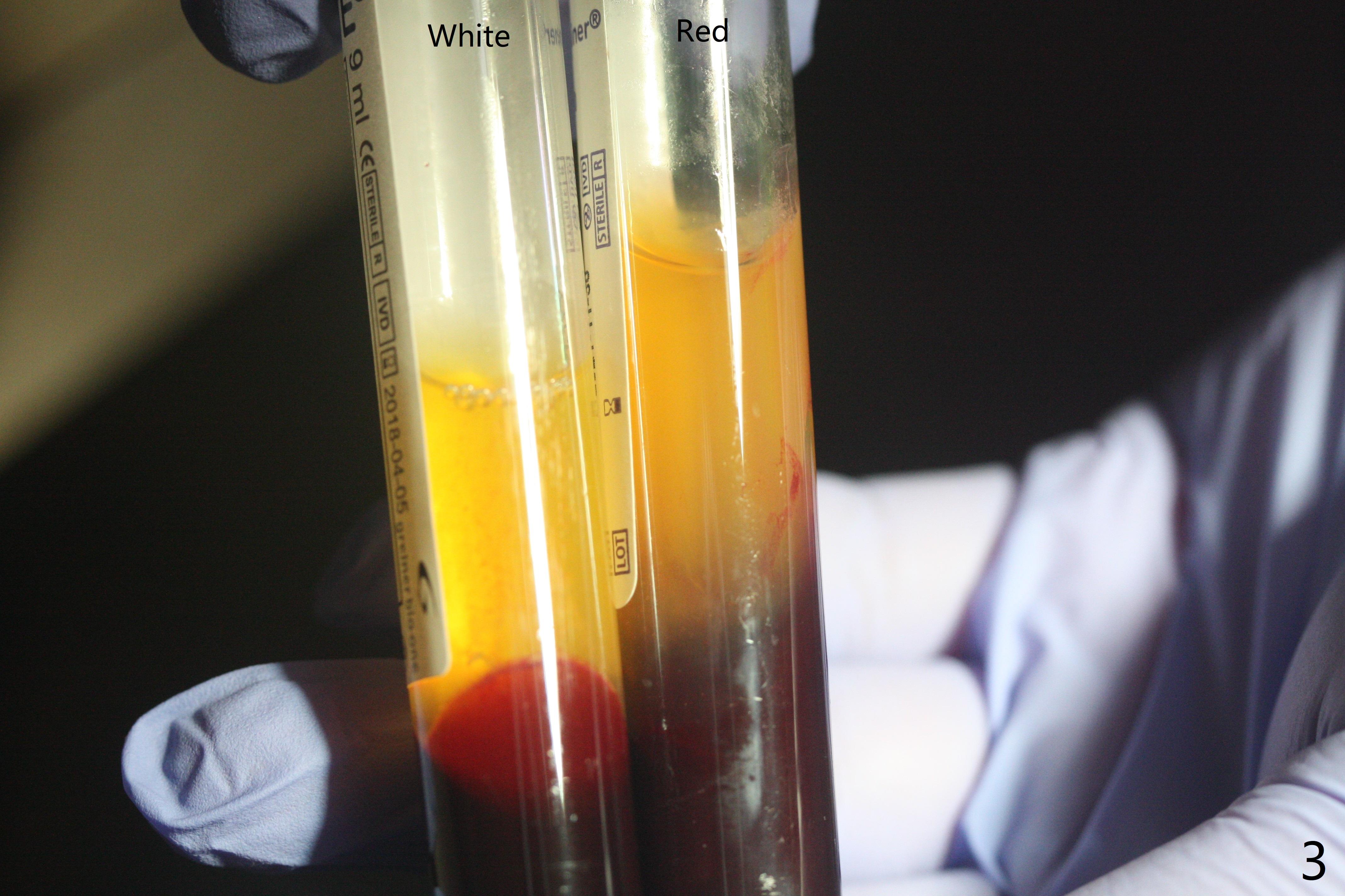
In spite of use of water pik, the coronal threads of the implant at #30 remain exposed 2 months postop (Fig.1). Envelop incision with mesial and distal accessory release ones reveals buccal bony defect (Fig.2). After 2nd spin (1500 RPM for 10 minutes), PRF forms in the red tube (Fig.3 yellow gel-like). Following use of Titanium brush, sticky bone is place (not so bone block-like, Fig.4), followed by a large piece of PRF membrane (from the red tube of Fig.3), Cytoplast (Fig.5,6 white porous) and a small piece of PRF membrane (from the white tube, next to the thin gingiva). The distal (Fig.6 D) and mesial (Fig.7 M) flaps are approximated (arrows without suture) as much as possible as well as lingual. 4-0 Polyglycolic Acid suture is used. Periodontal dressing dislodges 5 days postop. When the patient returns 1 week postop, he is pain free. Although Cytoplast is exposed, the surrounding gingiva seems to be healthy (Fig.8). Later the permanent crown of #31 is temporarily cemented with OHI. It appears that Cytoplast could be used to cover PRF membranes for soft tissue defect, followed by immediate provisional at the stage of immediate implant. The discolored (apparently contaminated) Cytoplast seems to be expelled 6 weeks postop (Fig.9). When the latter is removed, the soft tissue looks normal (Fig.10). Two weeks later, the apparently normal, but thin gingiva forms over the former granulation tissue (Fig.11 *, as compared to Fig.10). There is not enough bone coronal to the implant plateau 2 months post graft (Fig.12). Three months later, bone graft will be re-placed possibly with uncover.
Return to No Deviation Webinars 开场白 Xin Wei, DDS, PhD, MS 1st edition 07/15/2020, last revision 09/09/2020