,30%20Ncm,%20nearly%201%20y%20post%20cem,%20food%20impaction.jpg)
|
|
|
|
|
|
|
|
|
|
|
|
|
|
|
High Torque
M
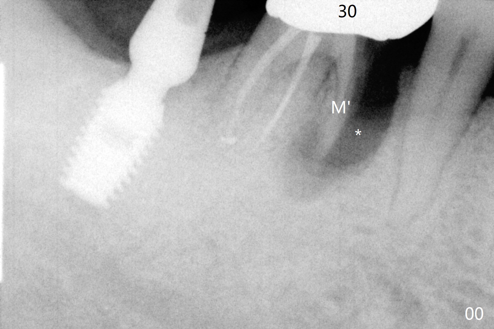
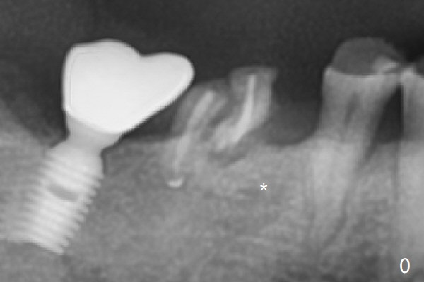
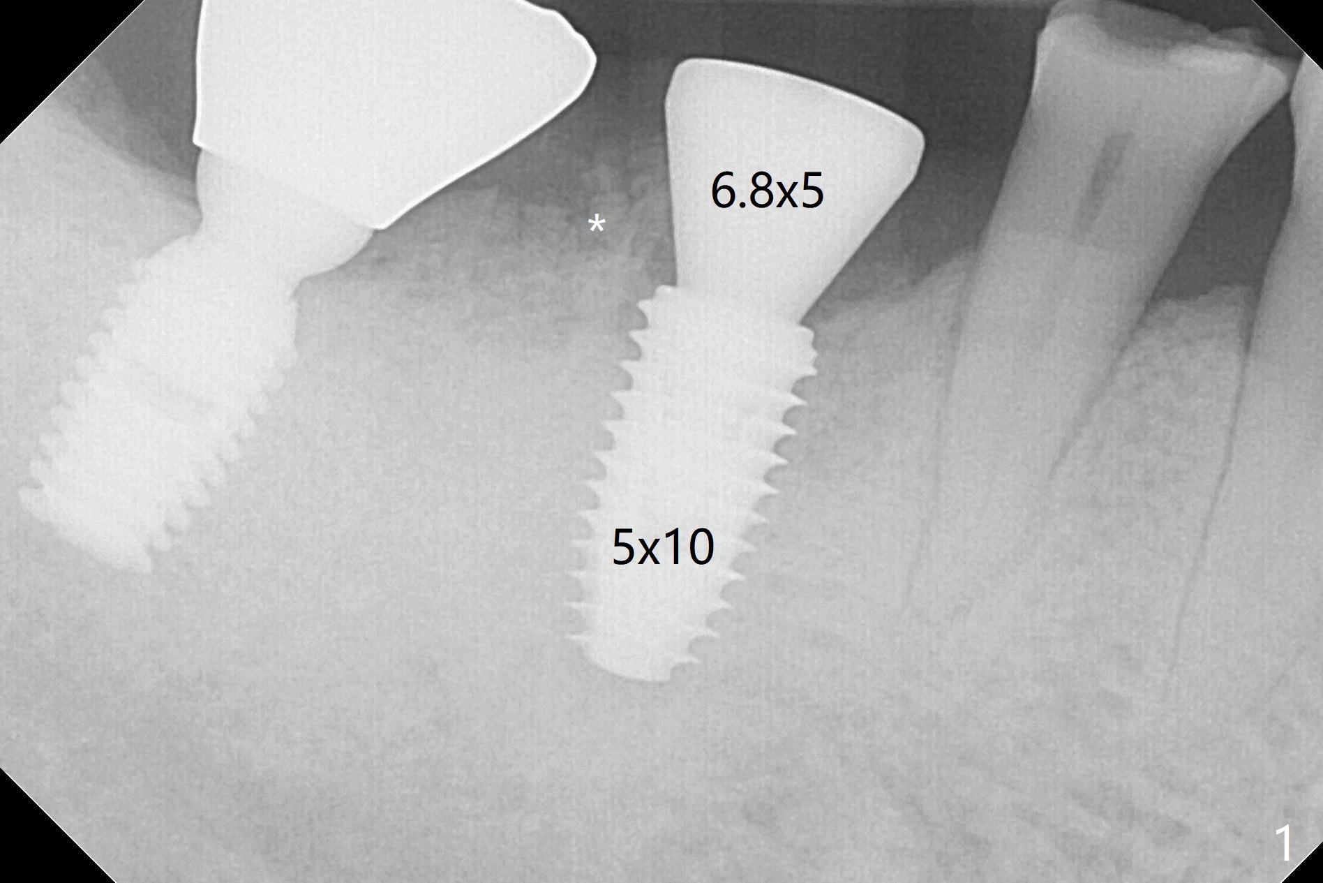
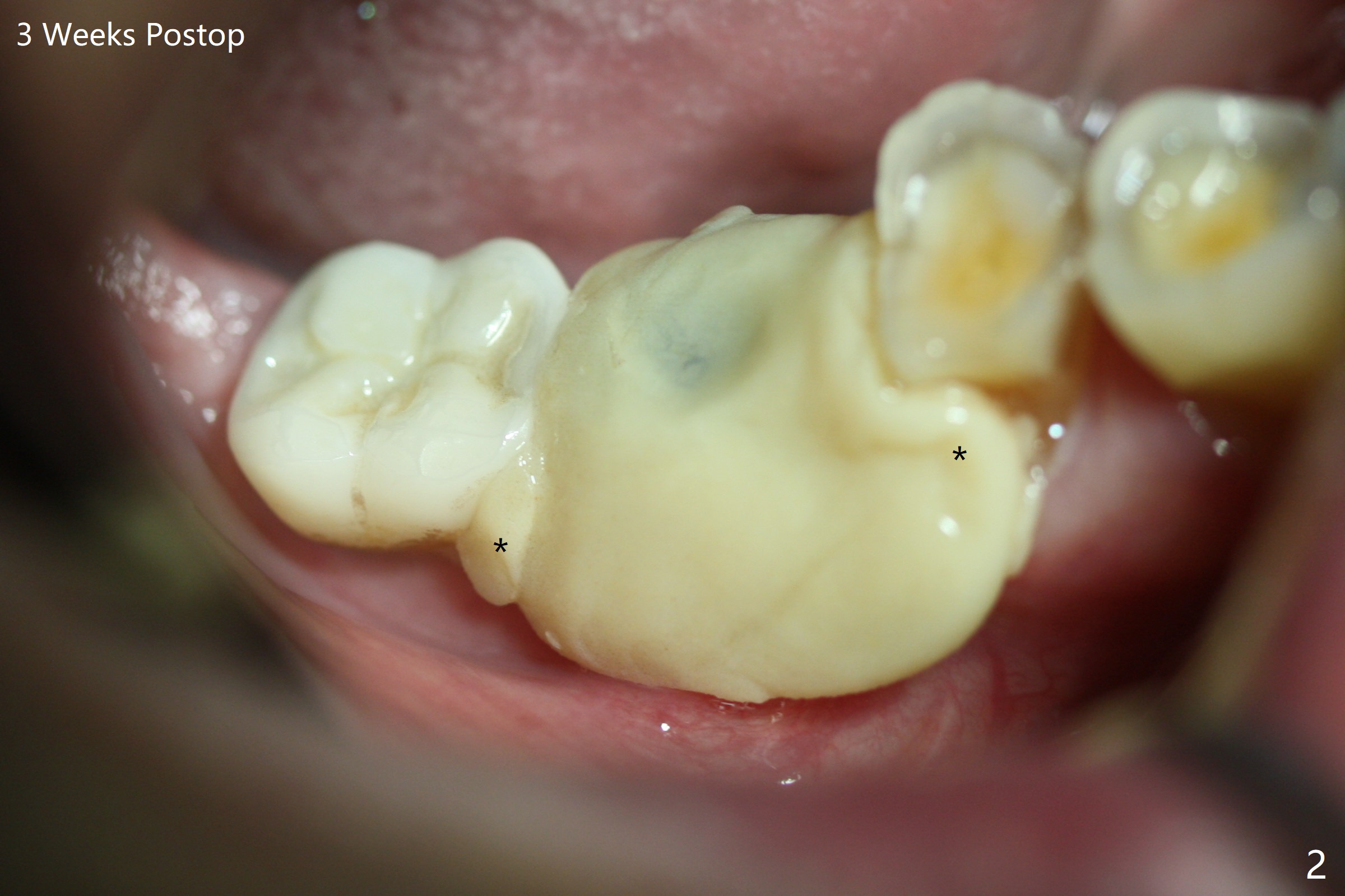
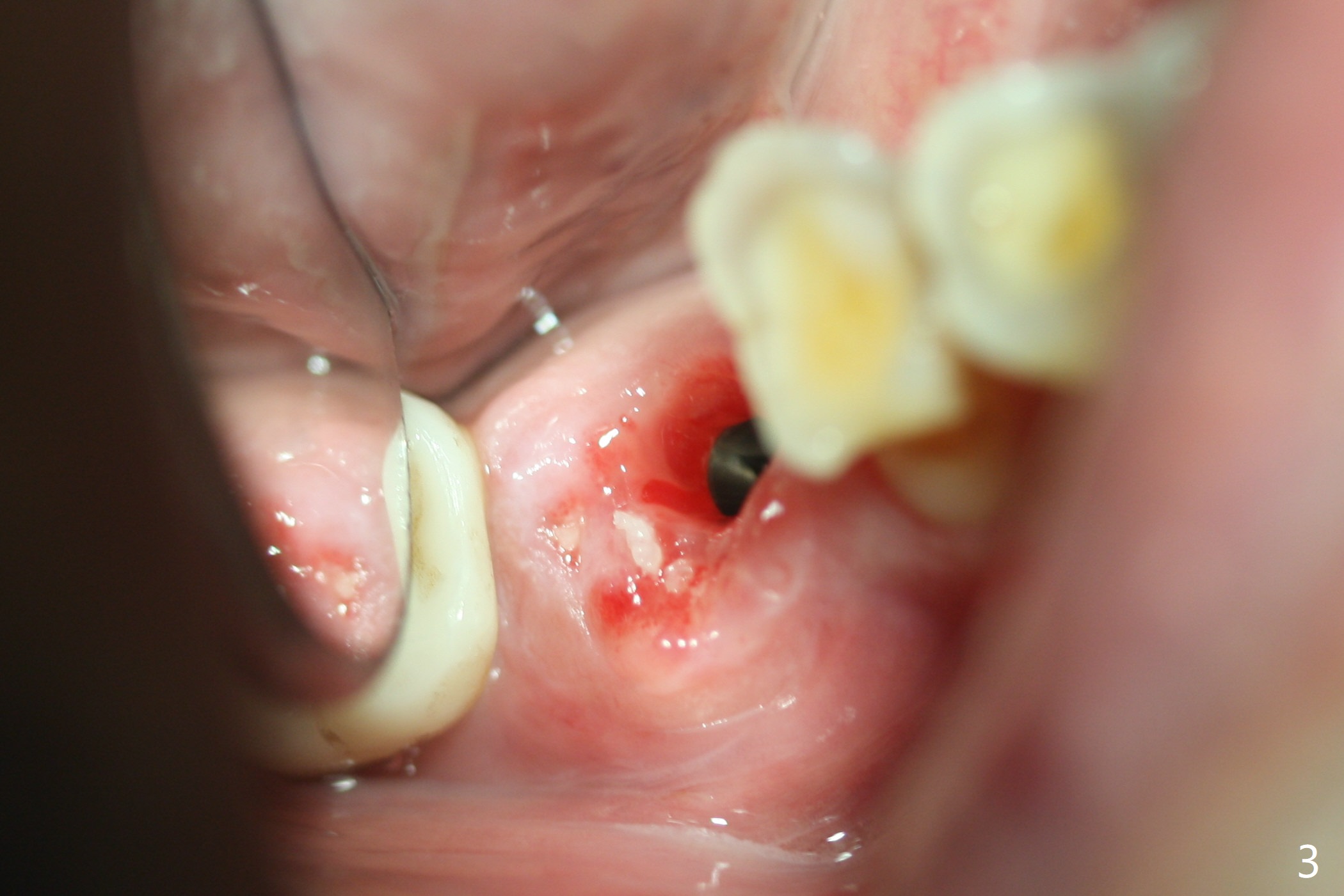
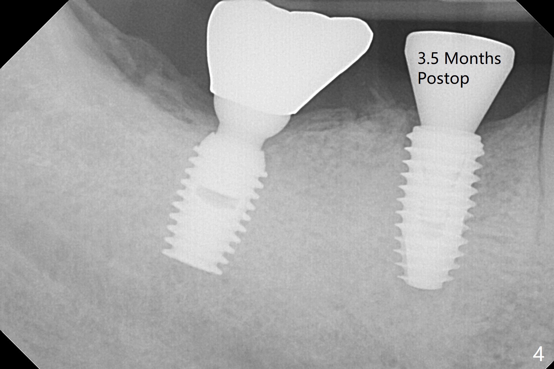
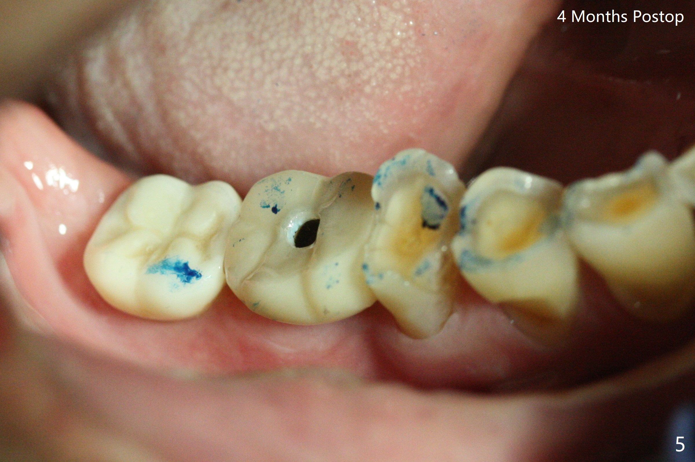
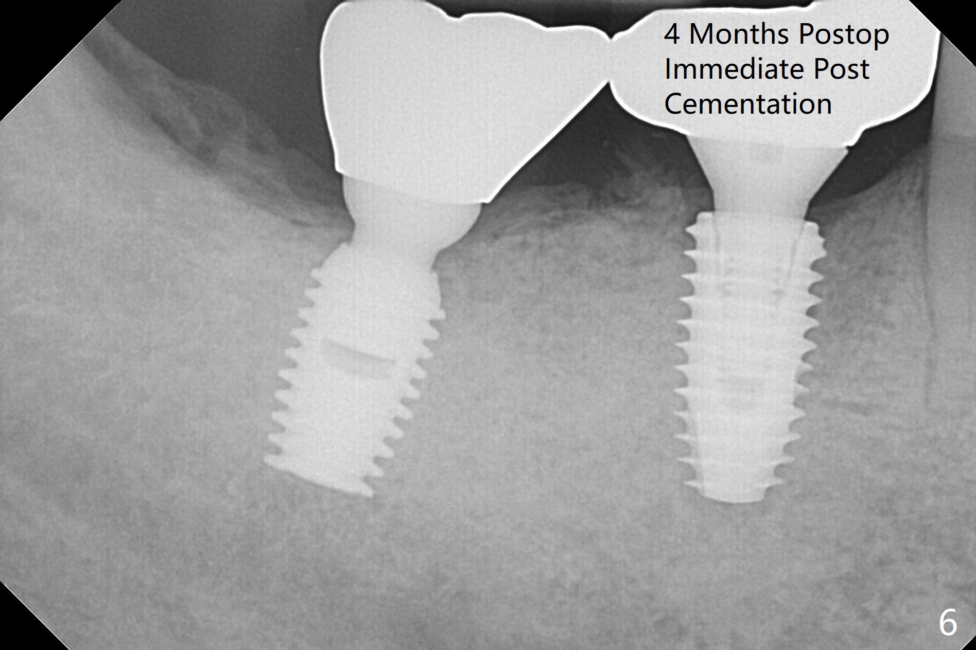
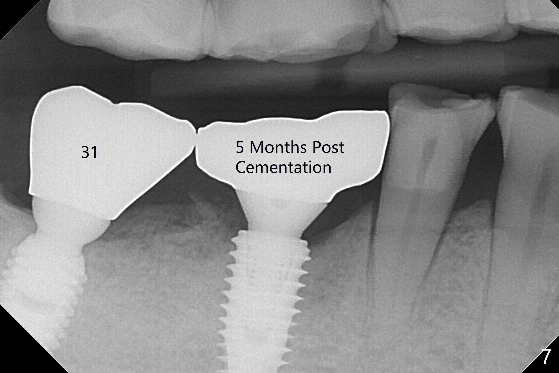
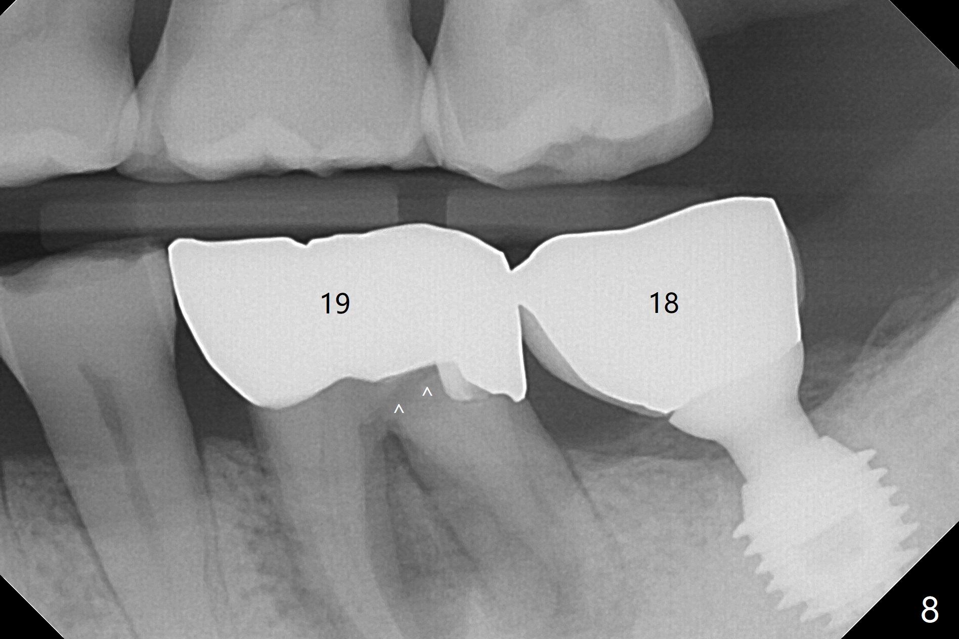
Vertical fracture of the mesial root of the tooth #30 after RCT is associated with bone loss (Fig.00 *). When the mesial portion of the mesial root (M', loose one) is exfoliated, the bone loss resolves (Fig.0). To reduce heat-induced bone necrosis at #19, osteotomy is conducted slowly with copious irrigation with cold saline. Bone density is felt while a 5x10 mm implant is being placed after using cortical tap to the 2nd line of the implant driver. The implant needs to be reverse torqued several times before reaching its final depth (Fig.1 (~50 Ncm)). Since the residual roots are superficially positioned, the immediate implant looks as a delayed one. Although the implant is placed mesial to the septum clinically, its position in X-ray seems to be normal. Because of severe wear and lack of vertical height, a 6.8x5 mm healing abutment is placed. Retention of bone graft (Fig.1 *) is maintained by spreading setting acrylic into the edentulous undercut areas (Fig.2 *). The so called "acrylic dressing" remains in place 3 weeks postop (Fig.8). When it is removed with the healing abutment, the wound heals (Fig.3). Note the limited vertical height. The bone graft placed in the distal socket appears to have been converted to the native bone 3.5 months postop (Fig.4). To reduce severe wear of the natural teeth, the occlusion of the new crown is not heavily decreased (Fig.5). It should be alright considering favorable crown/implant ratio (Fig.6). There is no bone loss 5 months post cementation, although the abutment screw is just retightened (Fig.7). In spite of poor trajectory associated with #18 (Fig.8) and 31 (Fig.7) Bicon implants, the abutments have not been dislodged. For the bruxer, the next implant at #19 with distal root fracture (Fig.8 ^) should be Bicon. The patient complains of food impaction nearly 1 year post cementation. The mesial and distal contacts of #30 crowns are light. When the abutment/crown is removed, there is implant well contamination (food debris). It appears that the previous abutment (5.7x4(2) mm, Fig.6,7) is incompletely seated. When a smaller abutment is placed and torqued at 30 Ncm, it is seated fully (Fig.9 (<: no gap)). New impression is taken. The distal gingival embrasure is larger than the mesial one because of the higher distal crest (Fig.6,7). If there is food impaction distal to the new crown, the distal crestal bone should be removed with lab closure of the embrasure.
Return to Lower Molar Immediate Implant, Prevent Molar Periimplantitis (Protocols, Table), Trajectory
Xin Wei, DDS, PhD, MS 1st edition 10/31/2018, last revision 02/22/2020